Figure 4. Representative fundus images of the same retina 14 days after injection of AAV-CMV-GFP. A is an image taken under bright light illumination. B represents the image with GFP signal only. Features in the retina are labeled as follows: BV = blood vessels, ONH = optic
nerve head, and RGCs = retinal ganglion cells. The images have been enhanced using the sharpen tool on Adobe Photoshop. Scale
bar = 200 μm.
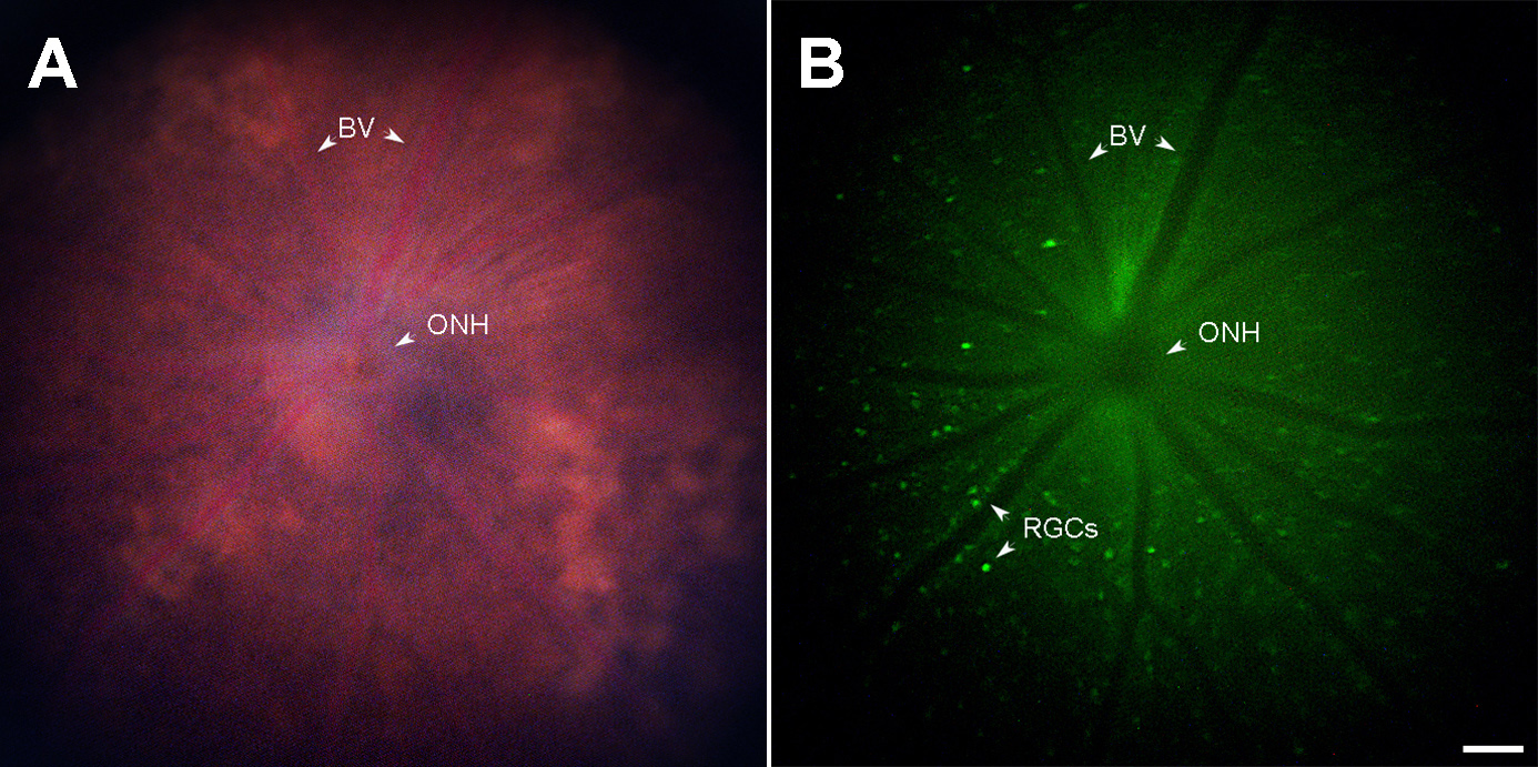
 Figure 4 of
Lin, Mol Vis 2025; 31:1-9.
Figure 4 of
Lin, Mol Vis 2025; 31:1-9.