Figure 3. Immunostained retinal flat mount. A: Representative retinal flat mount showing strong GFP expression throughout the entire retina. B: Some GFP-positive cells exhibit the dendritic morphology typical of RGCs and have an axon extending from the cells to the
optic disc, confirming their identity as RGCs. C–E: Immunostaining the retina with GFP, BRN3A, and POU6F2, indicating that virtually all the GFP-positive cells are RGCs. Scale
bar in A = 500 μm. Scale bar in B–E = 50 µm.
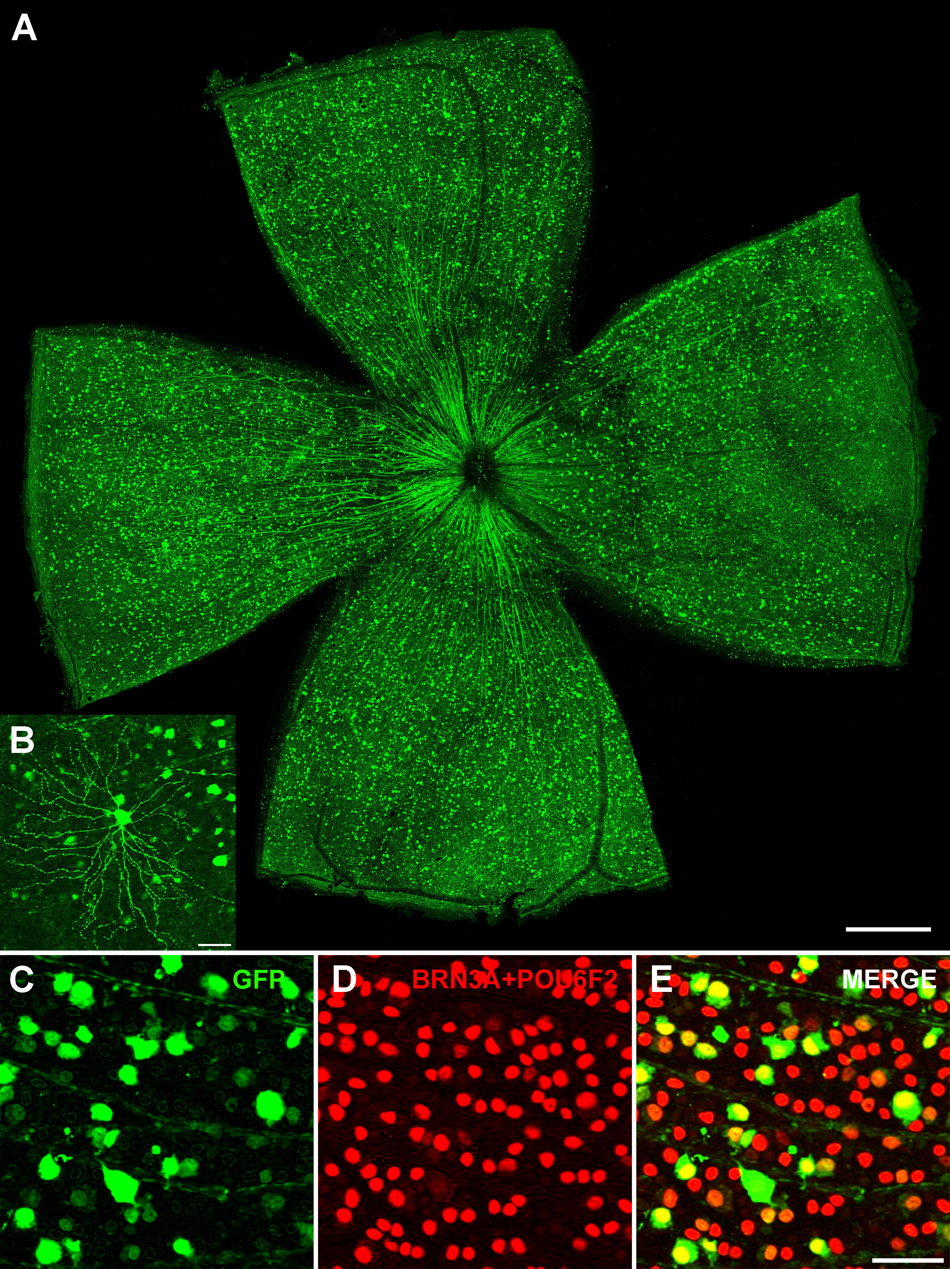
 Figure 3 of
Lin, Mol Vis 2025; 31:1-9.
Figure 3 of
Lin, Mol Vis 2025; 31:1-9.