Figure 1. Predicted tertiary structure of tissue inhibitor of metalloproteinases 3 (TIMP-3), showing residues mutated in Sorsby fundus
dystrophy (SFD). A model of human TIMP-3 was generated using AlphaFold and annotated using PyMOL. Residues located in the
signal peptide are indicated in purple, residues in the N-terminal domain are indicated in orange, and those in the C-terminal
region are indicated in blue. (A) depicts the ribbon structure of TIMP-3, and (B) depicts the surface of TIMP-3, showing surface residues mutated in SFD.
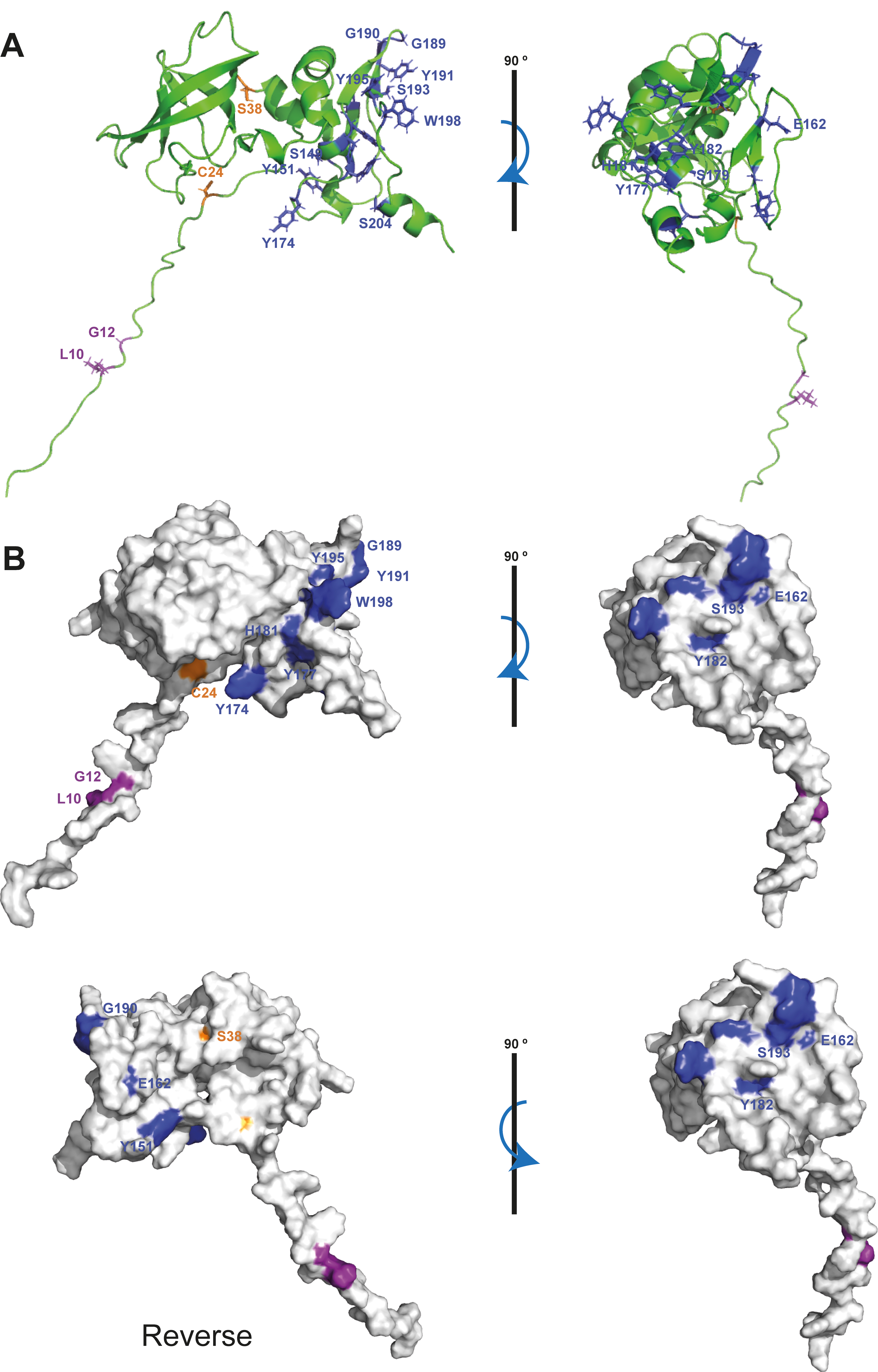
 Figure 1 of
Betts, Mol Vis 2024; 30:74-91.
Figure 1 of
Betts, Mol Vis 2024; 30:74-91.