Figure 1. Technique for standardizing cell density measurement using ImageJ. A: On the left is the mosaic image of the damaged lens with the five counting zones, which are positioned thanks to the macro.
On the right is the image of the corresponding healthy lens with the five zones automatically positioned at the same locations.
At the bottom is an example of zone four of each image. B: Automatic counting of Hoechst-stained nuclei with the CorneaJ plugin.
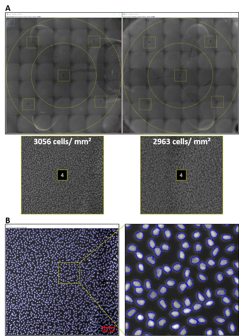
 Figure 1 of
Poinard, Mol Vis 2024; 30:478-487.
Figure 1 of
Poinard, Mol Vis 2024; 30:478-487.