Figure 2. Clinical features of Patient 2. A, B: Systematic examination revealed a protuberant umbilicus. C: Dental examination revealed widely spaced teeth as well as some abnormally shaped teeth. D, E: Slit-lamp images showed posterior embryotoxon (black arrowhead), corectopia, and iris hypoplasia in both eyes. F, G: UBM revealed high insertion of the iris root and iris anterior synechia. H, I: Scanning laser ophthalmoscopey (SLO) indicated that the cup-to-disk ratios were 0.3 (OD) and 0.4 (OS).
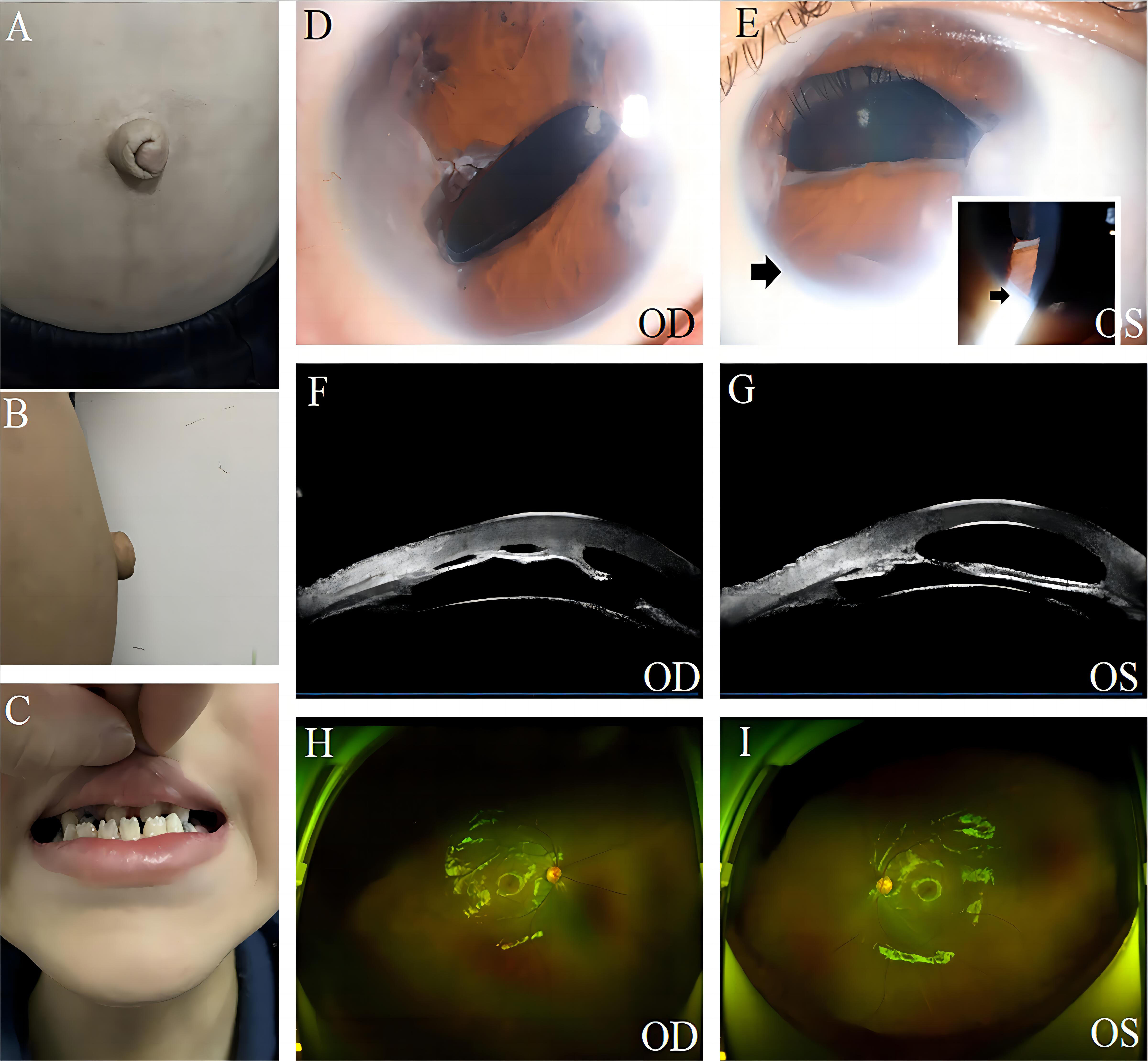
 Figure 2 of
Jiang, Mol Vis 2024; 30:466-476.
Figure 2 of
Jiang, Mol Vis 2024; 30:466-476.