Figure 7. Smad3 directly associated with stromal ECM protein modulation impacted the corneal stromal wound healing in mice after alkali
injury during active wound healing. A: H&E staining showing the morphological architecture of Smad3+/+ naïve (a); Smad3−/− naïve (b); Smad3+/+ post-alkali injury (c); and Smad3−/−- post-alkali injury (d) corneal tissue sections at day 21. No structural changes were observed in Smad3−/− and Smad3+/+ naïve corneal tissue sections. An increased cellular infiltration and distorted collagen lamellae (arrows) were observed
in the corneal stroma of Smad3+/+ and Smad3−/− compared with Smad3−/− mouse corneal tissue sections after alkali injury at day 21. B: Masson’s trichrome staining showed the gross collagen level in Smad3+/+ naïve (a); Smad3−/− naïve (b); Smad3+/+ post-alkali injury (c); and Smad3−/− post-alkali injury (d) corneal tissue sections at day 21. No collagen levels were altered in Smad3−/− and Smad3+/+ naïve corneal tissue sections. Increased collagen deposition was observed in the corneal stroma of Smad3+/+ compared with Smad3−/− mouse corneal tissue sections after alkali injury at day 21, as indicated by increased blue color intensity (arrowhead).
Scale bar = 200 μm.
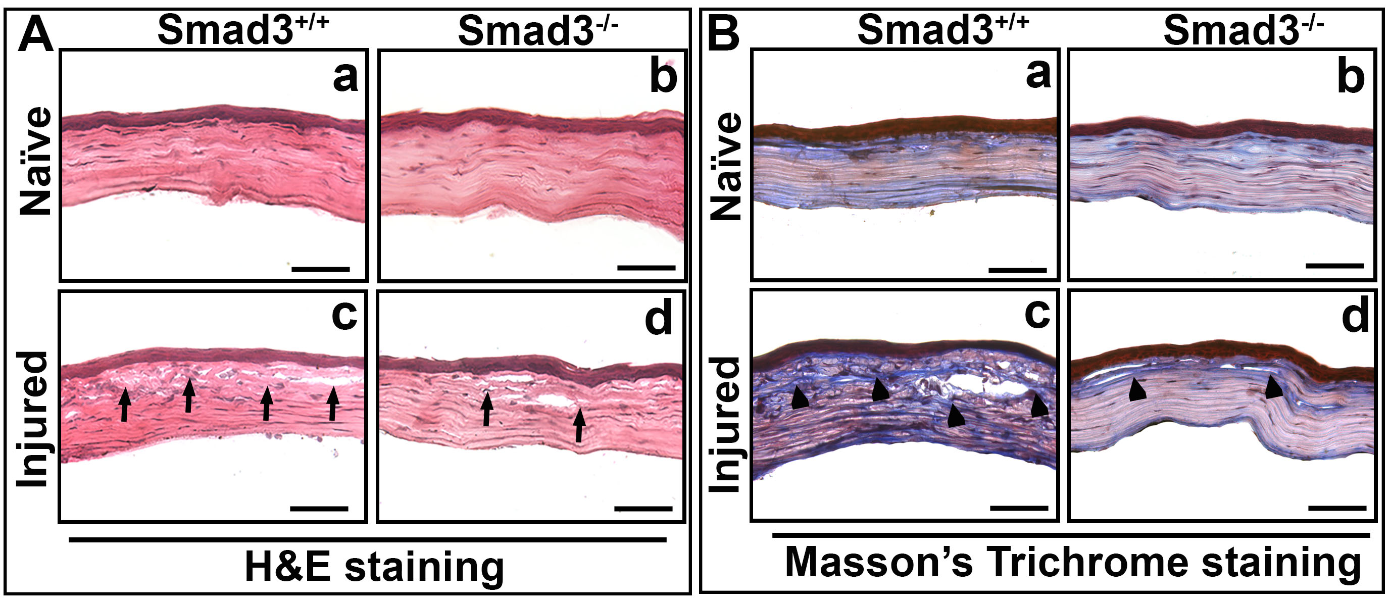
 Figure 7 of
Gupta, Mol Vis 2024; 30:448-464.
Figure 7 of
Gupta, Mol Vis 2024; 30:448-464.