Figure 6. Smad3 gene deficiency regulates the stromal ECM protein modulation during fibrosis in mice after alkali injury. A: The comparative mRNA expression of α-SMA (fibrosis marker) was significantly downregulated in Smad3−/− mouse corneal tissue compared with Smad3+/+ mouse corneal tissue after alkali injury. B: The immunofluorescence staining of α-SMA was prominently reduced in Smad3−/− mouse corneal tissue compared with Smad3+/+ mouse corneal tissue after alkali injury, whereas no changes were recorded in both Smad3+/+ and Smad3−/− mouse naïve corneal tissue sections. C: The western blot images showed that the α-SMA protein level was notably abridged in Smad3−/− mouse corneal tissue compared with Smad3+/+ mouse corneal tissue after alkali injury, whereas no changes were recorded in both Smad3+/+ and Smad3−/− mouse naïve corneal tissue sections. D: The western blot densitometry analysis graph shows a significant reduction in fibrosis level in Smad3−/− mouse corneas compared with Smad3+/+ mouse corneas. The results are expressed as mean ± SEM, and p<0.05 was considered significant. Scale bar = 200 µm.
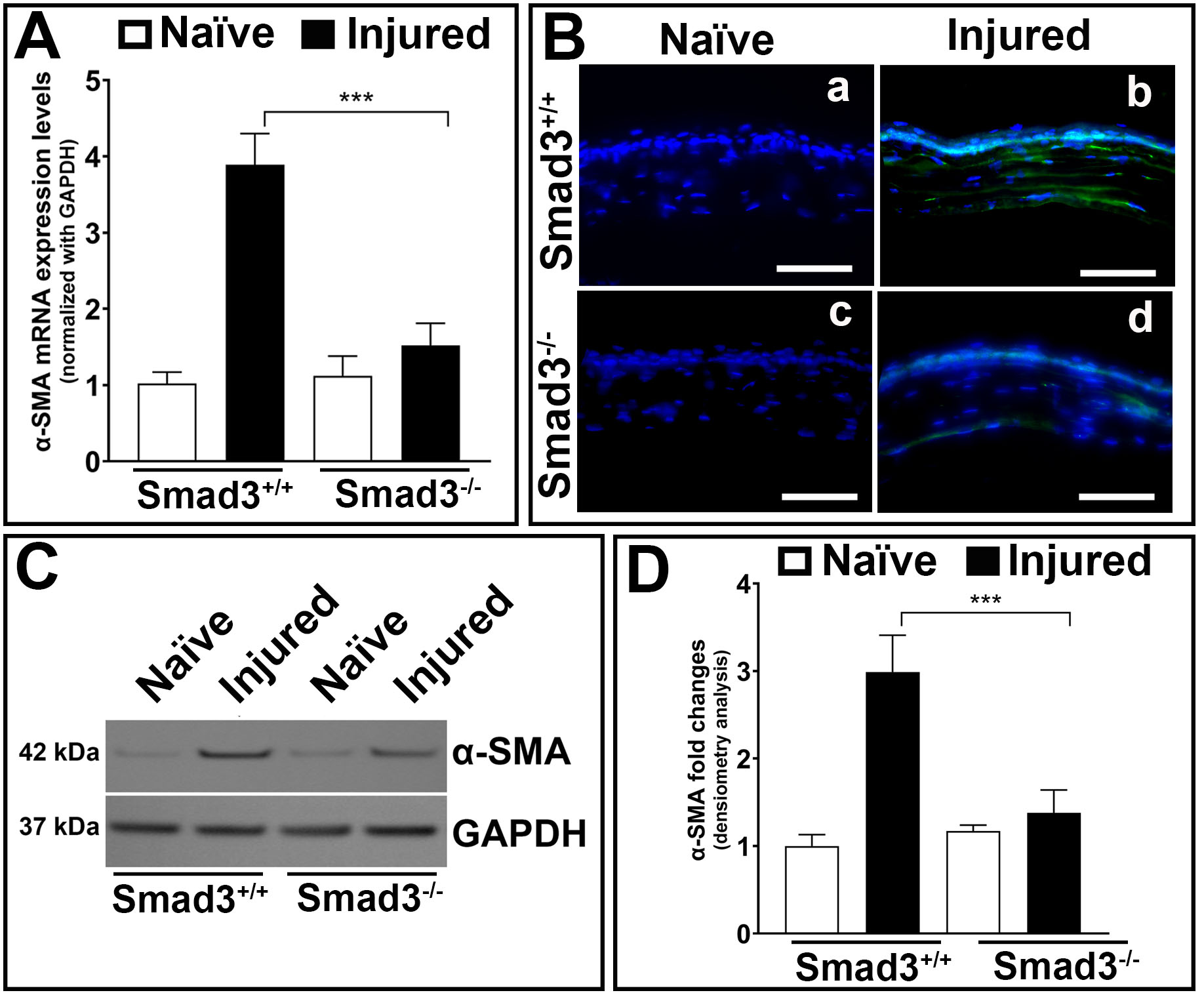
 Figure 6 of
Gupta, Mol Vis 2024; 30:448-464.
Figure 6 of
Gupta, Mol Vis 2024; 30:448-464.