Figure 2. Smad3 gene deficiency reduced corneal haze in mice after alkali injury. The representative stereomicroscopic mouse corneal
tissue images show the A: naïve; B: day 3; C: day 7; and D: day 21 post-alkali haze formation in Smad3+/+ mouse strain corneal tissue. The stereomicroscopic mouse corneal tissue images show the E: naïve; F: day 3; G: day 7; and H: day 21 post-alkali haze formation in Smad3−/− mouse strain corneal tissue. I: The haze quantification graph shows that corneal haze was significantly reduced in Smad3−/− compared with Smad3+/+ mouse corneal tissue (**p<0.01, ***p<0.001, scale bar = 0.5 mm).
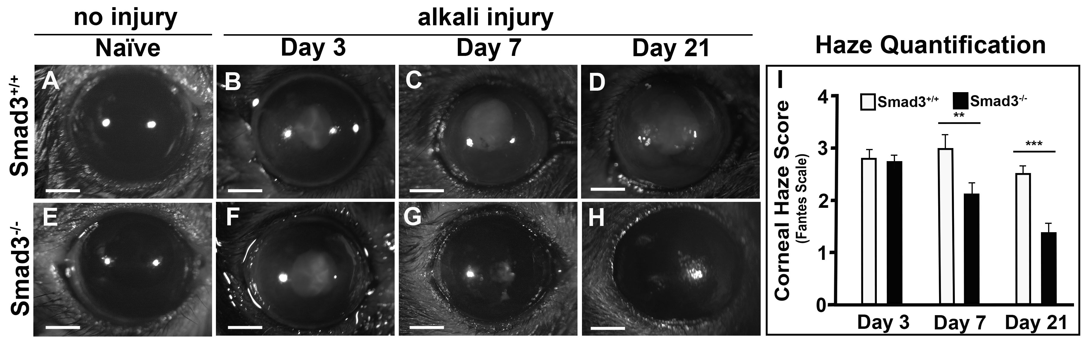
 Figure 2 of
Gupta, Mol Vis 2024; 30:448-464.
Figure 2 of
Gupta, Mol Vis 2024; 30:448-464.