 Appendix 3 of
Chacon-Camacho, Mol Vis 2024; 30:400-408.
Appendix 3 of
Chacon-Camacho, Mol Vis 2024; 30:400-408. Appendix 3 of
Chacon-Camacho, Mol Vis 2024; 30:400-408.
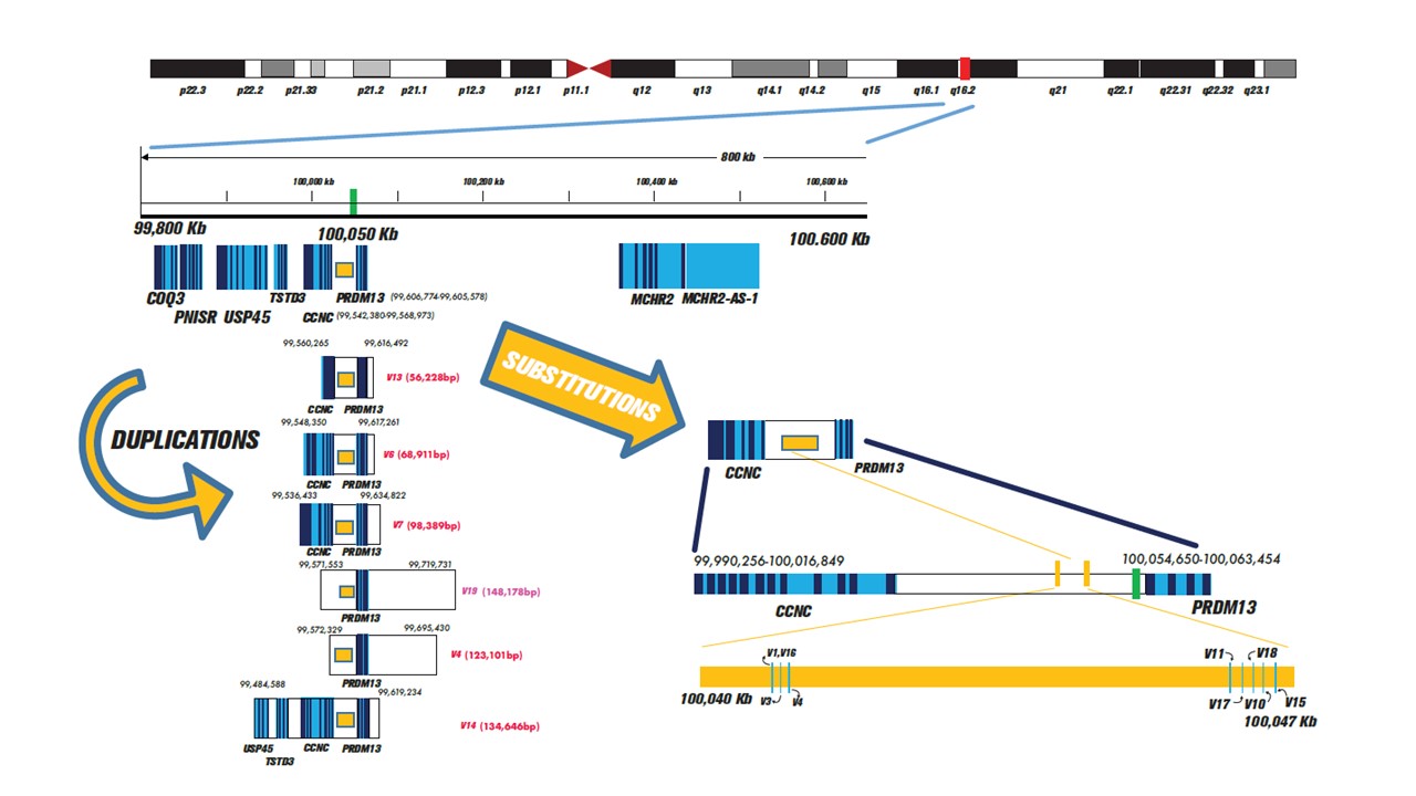
Appendix 3. Supplementary Figure 3.
To access the data, click or select the words “Appendix 3.” NCMD causing variants identified to date. A partial chromosome 6 view is shown in the upper part of the figure. MCDR1 locus (6q16.2) is delimited in a red box. Below, there is a zoom of the 6q16.2 region spanning approximately 800 kb. The light blue regions of each gene represent the introns and the vertical lines within each gene represent the exons of the gene; thicker vertical lines represent multiple exons. The white regions between the genes represent the intergenic regions, the most important of which is the one found between the CCNC and PRDM13 genes, which is called the DNA I hypersensitive region. Within this, NCMD-causing SNVs are located (yellow rectangle), in a region covering approximately 6 kb (enlarged on the right side of the image). On the left side of the image, all MCDR1 duplications reported on the date, including their genomic coordinates and their length are presented. The white boxes in the duplications represent intergenic regions. The novel duplication reported in the present work is indicated as V19.