Figure 2. Representative images of corneal sections for histochemistry (HC-AB staining) and immunohistochemistry (IHC-5D4 MoAb immunoreactivity)
analysis of normal (control) and MCD patients with different mutation types. Images on the left side represent AB-stained
corneal sections, and the right side represents immunostained sections. A: The corneal layers marked in the control (cadaver) show normal morphology. B–F represent images of unrelated MCD patients (Patients 7, 17, 29, 60–1, and 67) harboring different CHST6 mutations. Major
changes were observed for the patients harboring deletion (del), deletion-insertion (delins), and deletion of ORF mutations
with severe morphological alterations in the BL and anterior stroma due to abnormal, amorphous, finely granulated GAG deposits
(*) with altered collagen lamellae and altered keratocyte (K) cell shape. IHC also showed the presence of KS only in the stroma,
with a smaller number of stromal keratocytes. F represents the images for the MCD patient with no coding region CHST6 mutation, showing a normal corneal morphology like
the control (A). In the IHC images, red represents PI staining, and green indicates FITC-labeled anti-KS antibodies. Ep, epithelium; BL,
Bowman’s layer; BM, basement membrane; S, stroma; DM, Descemet’s membrane; EN, endothelium; K, keratocytes.
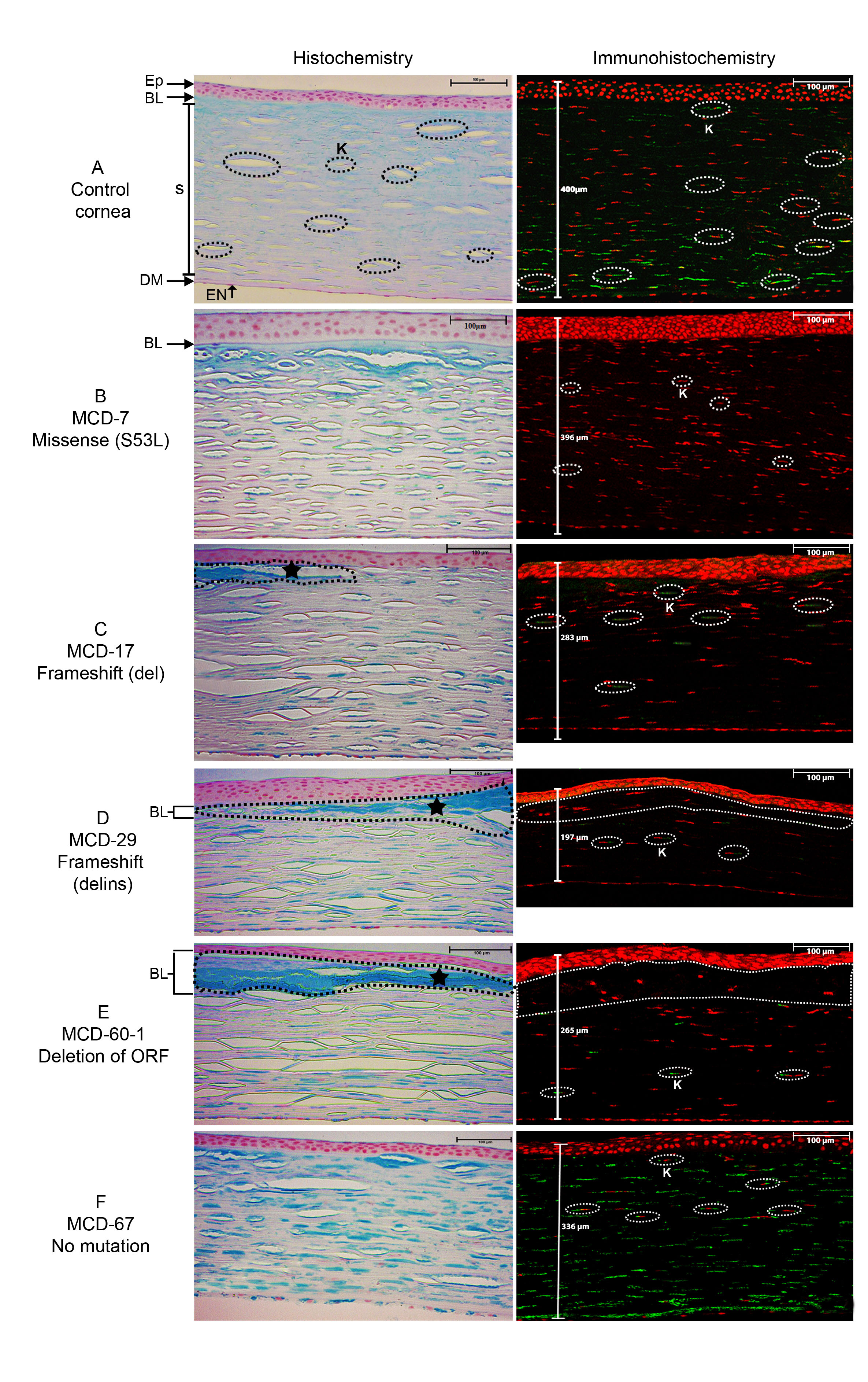
 Figure 2 of
Murugan, Mol Vis 2024; 30:305-318.
Figure 2 of
Murugan, Mol Vis 2024; 30:305-318.