Figure 1. Fluorescein angiography of experimental CRVO. A–B: Venous tortuosity and dilation, and flame-shaped hemorrhages appearing within 30 min after induced CRVO. Experimental CRVO
was treated with either ranibizumab or NaCl (control). C–D: Flame-shaped hemorrhages in the peripheral retina following experimental CRVO.
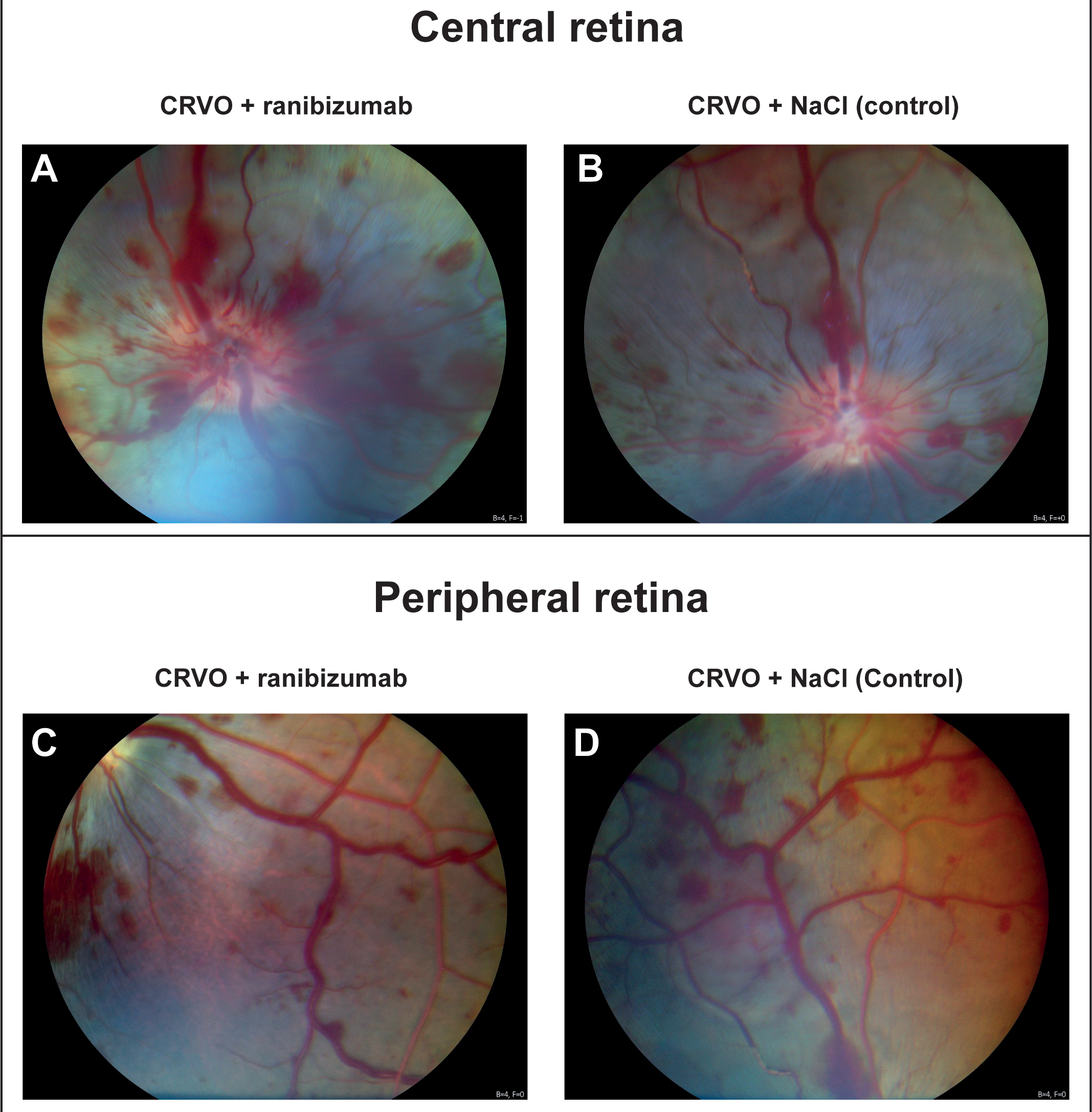
 Figure 1 of
Cehofski, Mol Vis 2024; 30:268-277.
Figure 1 of
Cehofski, Mol Vis 2024; 30:268-277.