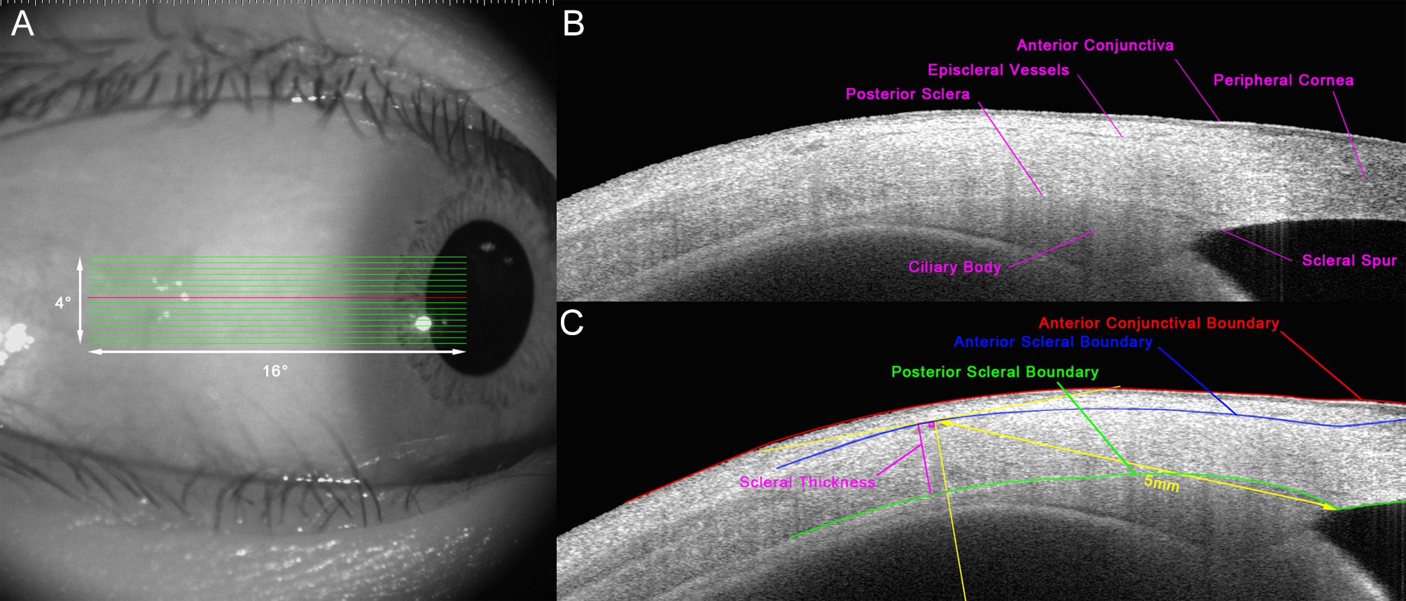
Figure 1. AS-OCT images.s A: AS-OCT image of the anterior scleral scanning protocol. A 16° × 4° volume B-scan with 21 lines was conducted to obtain images
of the anterior temporal sclera. B: A B-scan of the anterior temporal sclera with anatomic landmarks labeled. C: A B-scan with semi-automated segmentation, delineating the anterior scleral boundary (marked in blue), the posterior scleral
boundary (marked in green), the anterior conjunctiva boundary (marked in red), and the scleral thickness (marked in amaranth).

 Figure 1 of
Li, Mol Vis 2024; 30:229-238.
  Figure 1 of
Li, Mol Vis 2024; 30:229-238.