Figure 4. Comparison of the examination results of the macula of the right eye (No. 19: CM). A: Color fundus image, (B) enlarged image of white frame in A, (C) OCT image, (D) enlarged image of white frame in C. At the position where WDs (black circle) are seen in B in the SLO fundus image, hyperreflective lesions (red circles) in the RPE layer are seen in the OCT image (C and D). The black dashed line represents the cutting plane of OCT. E–H: HE-stained sections in and around the fovea. (I, J) Electron microscopic sections of RPE in H. Arrowheads in I indicate vacuolization with microgranules in the RPE cell. F–H: Enlarged image of three arrowheads in E. J: Enlarged image of arrowhead in I. Structures resembling lysosomes (arrowheads) are present in the cytoplasm; these vacuoles are found only in the macula.
Scale bars: 200 μm in A–D; 100 μm in E; 20 μm in F–I; 5 μm in J.
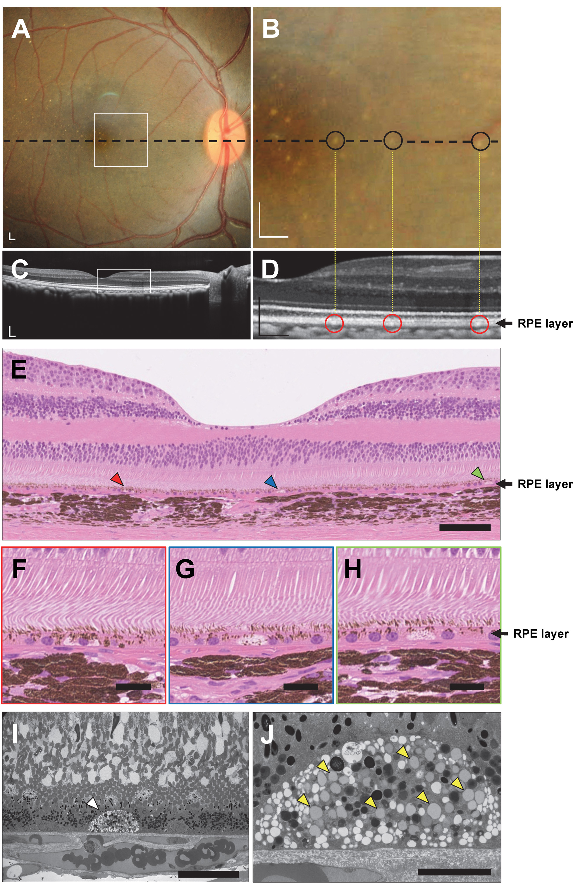
 Figure 4 of
Araki, Mol Vis 2024; 30:219-233.
Figure 4 of
Araki, Mol Vis 2024; 30:219-233.