Figure 2. Representative fundus images of CMs and RMs with WDs in the macula. (A) Normal (No. 7: CM, 6 years old), (B) Normal (No. 29: RM, 5 years old), (C) WDs in the macula (No. 19: CM, 11 years old), (D) WDs in the macula (No. 51: RM, 14 years old). The normal fovea is a dark portion present on the temporal side shown in A and B. Many granular WDs (arrowheads) are concentrated in and around the fovea in C and D. E–M: Representative WDs in and around the fovea in CMs (Nos. 17, 18, and 20 from the upper left; Nos. 21, 22, and 23 from the
middle left; and Nos. 24, 25, and 26 from the lower left). N–V: Representative WDs in and around the fovea in RMs (Nos. 43, 45, and 46 from the upper left; Nos. 47, 48, and 49 from the
middle left; and Nos. 50, 52, and 53 from the lower left). Many granular WDs are concentrated in and around the fovea. Some
animals have WDs throughout the macula. The number of WDs across individuals is varied. However, almost all WDs have similar
shapes, size, and color in both left and right eyes, and no significant difference in these characteristics are observed between
CMs and RMs. Scale bars: 500 μm in A–D; 250 μm in E–V.
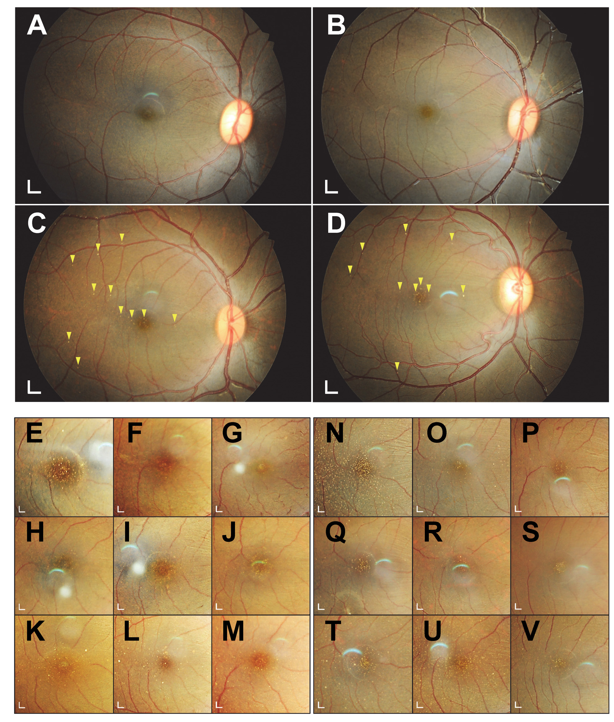
 Figure 2 of
Araki, Mol Vis 2024; 30:219-233.
Figure 2 of
Araki, Mol Vis 2024; 30:219-233.