Figure 4. SFN suppresses the formation of stress fibers induced by TGF-β in HTFs. (A) After embedding with collagen gels, cells were
incubated for 24 h with or without SFN, followed by a 24 h culture with or without TGF-β. The formation of stress fibers was
determined using Alexa Fluor 568–labeled phalloidin and DAPI staining. Scale bars: 50 µm. Fluorescence intensity analysis
in (A) is shown in (B). Data are presented as mean ± SD (n=3). *p<0.05 compared to controls. †p<0.05 compared to cells treated
with TGF-β alone.
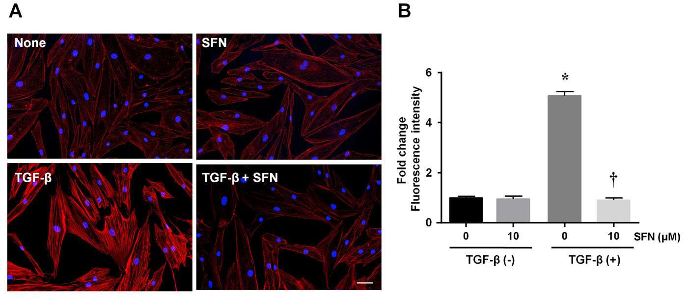
 Figure 4 of
Liu, Mol Vis 2024; 30:200-210.
Figure 4 of
Liu, Mol Vis 2024; 30:200-210.