Figure 8. Histopathological examination examples of Group 1 (A), Group 2 (B), Group 3 (C), and Group 4 (D). Red arrows indicate the
newly formed epithelial layers. Black arrows indicate the degree of stromal collogen remodeling and keratocyte loss. Blue
arrows indicate the newly formed vessels, and blue stars indicate PMNL infiltration (HE; x200). Abbreviations: PMNL, polymorphonuclear
leukocyte.
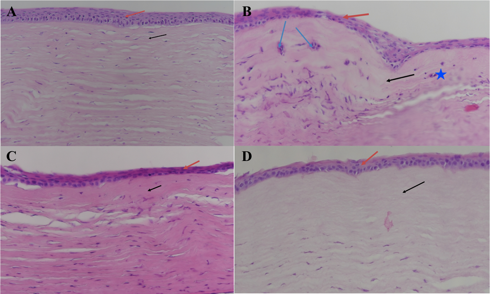
 Figure 8 of
Korkmaz, Mol Vis 2024; 30:188-199.
Figure 8 of
Korkmaz, Mol Vis 2024; 30:188-199.