Figure 2. Protocol of longitudinal IVCM monitoring of dry eye development. A: Modified HRT3 platform for animal research. B: The flowchart of longitudinal IVCM monitoring of dry eye neural changes. Pre-surgery: guide image of the posterior stromal
nerve was recorded, and corresponding SNP images were recorded anteriorly through volume acquisition. Post-surgery: The same
posterior stromal nerve was located after DLGR, and SNP images were recorded and compared to previous SNP images. Note that
the similar morphologies of stromal nerves of guide image and SNPs illustrate that the same corneal area was imaged at different
time points. SNP nerves were counted (white/yellow lines with white/yellow arrows), as well as cell infiltration (white/purple
dots with white/purple triangle arrows). C: A typical set of IVCM images before and after surgery, showing similar morphology to guide images and SNPs. White lines
and/or white arrows: SNP nerves detected in both pre- and post-surgery SNPs. Yellow lines and/or yellow arrows: SNP nerves
detected only in post-surgery SNPs. White dots and/or white triangles: SNP cells detected in both pre- and post-surgery SNPs.
Purple dots and/or purple triangle arrows: SNP cells detected only in post-surgery SNPs.
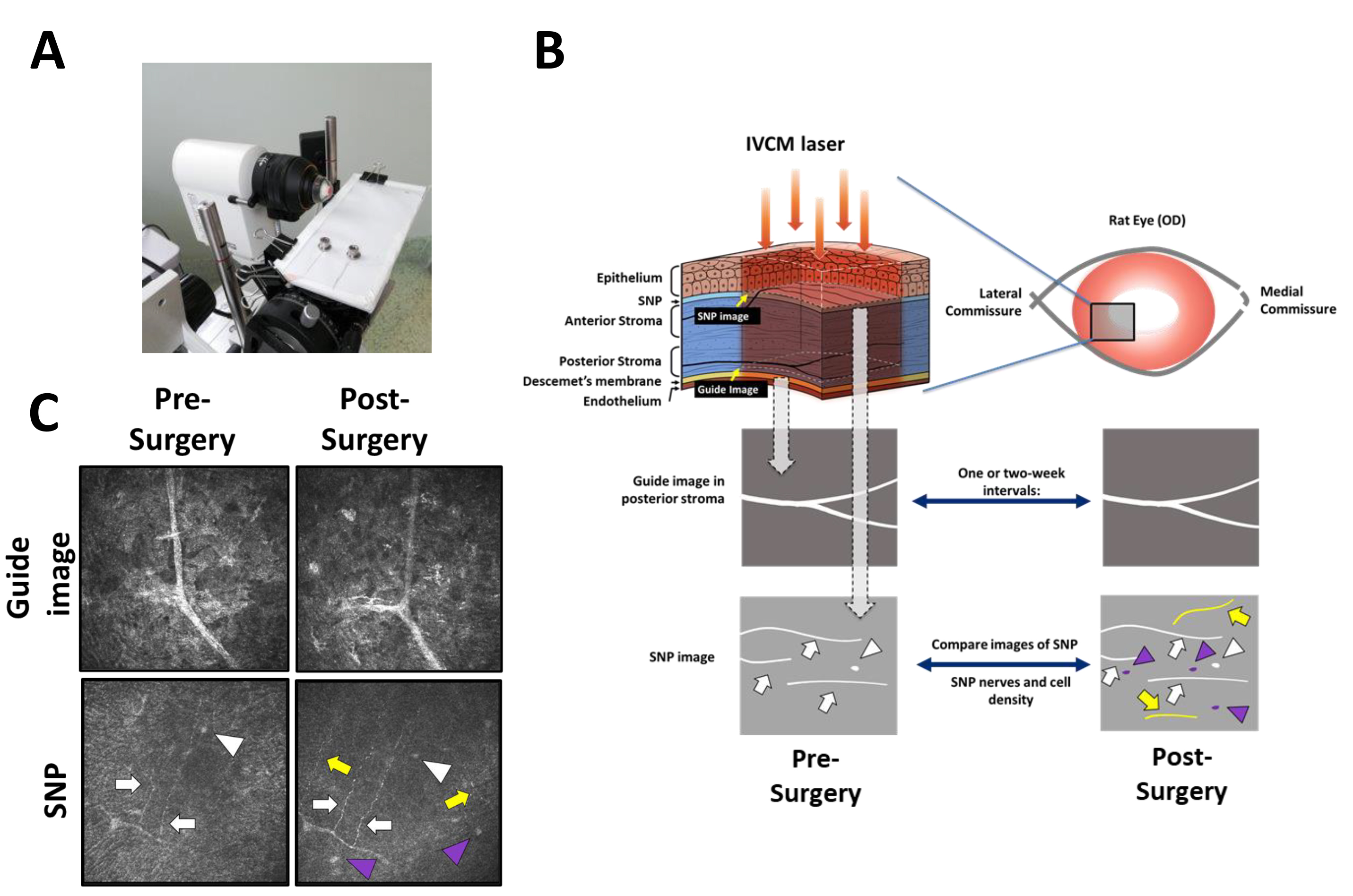
 Figure 2 of
Chen, Mol Vis 2024; 30:150-159.
Figure 2 of
Chen, Mol Vis 2024; 30:150-159.