Figure 5. Colocalization and coimmunoprecipitation of OPTN-GFP with Hsp72 in HTM cells. A: Cells transduced with an adenovirus expressing optineurin-GFP were counterstained with DAPI, and Hsp72 (red) was visualized
with a Cy-3-conjugated secondary antibody. Two panels represent the same cells. B: Cell lysate made from cells transduced with an adenovirus expressing OPTN-GFP was immunoprecipitated with an anti-GFP antibody
and then probed with an anti-Hsp72 antibody and vice versa. 1, input; 2, immunoprecipitation with resin only; 3, immunoprecipitation
with resin plus the indicated antibodies.
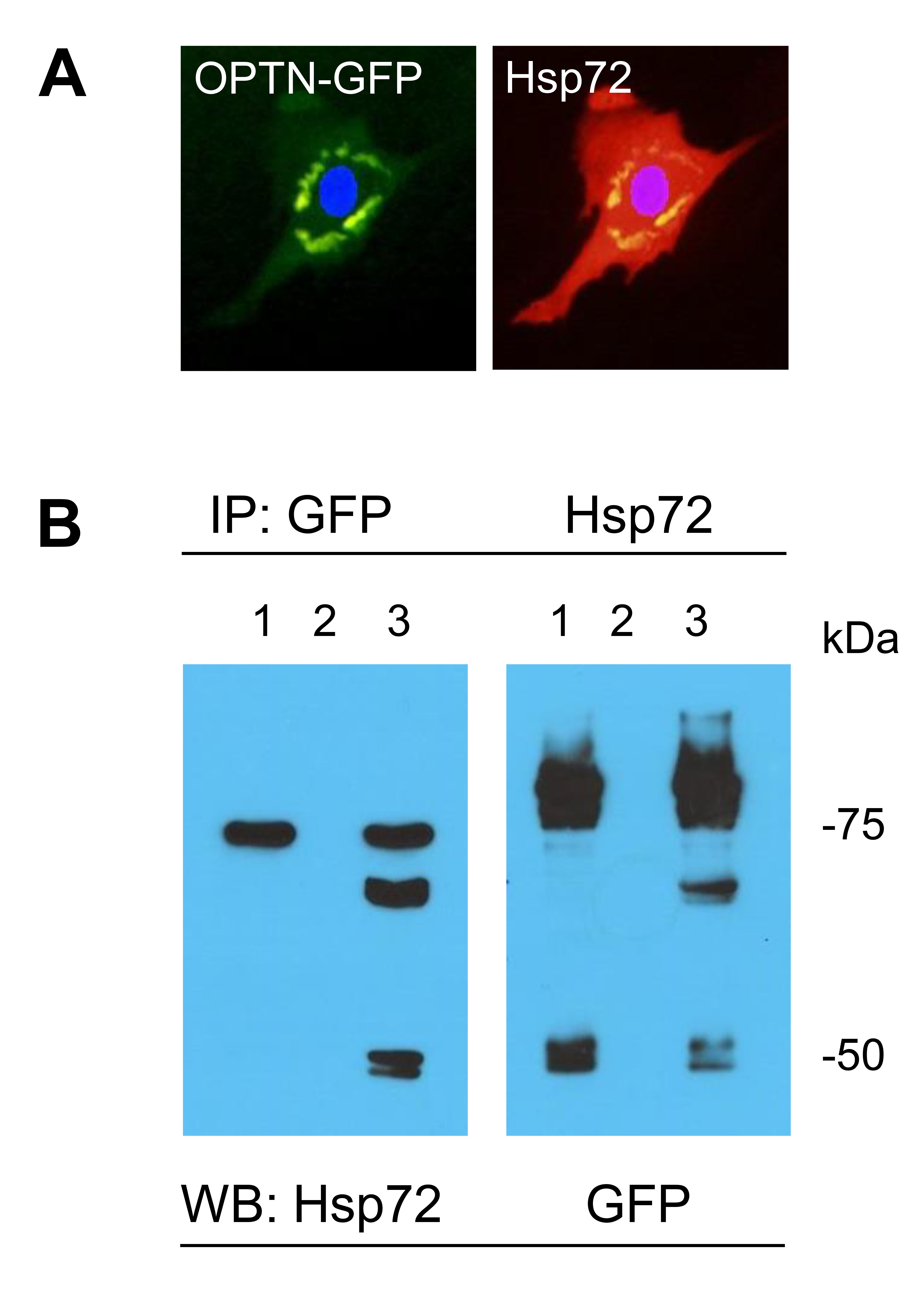
 Figure 5 of
Suh, Mol Vis 2024; 30:114-122.
Figure 5 of
Suh, Mol Vis 2024; 30:114-122.