Figure 4. Typical images of OPTN-GFP against the contour of HTM cells. Merged images for phase-contrast and green fluorescence were
photographed from the same cells transduced with adenoviruses expressing the indicated proteins. All image acquisition was
made with commonly available FITC filter set.
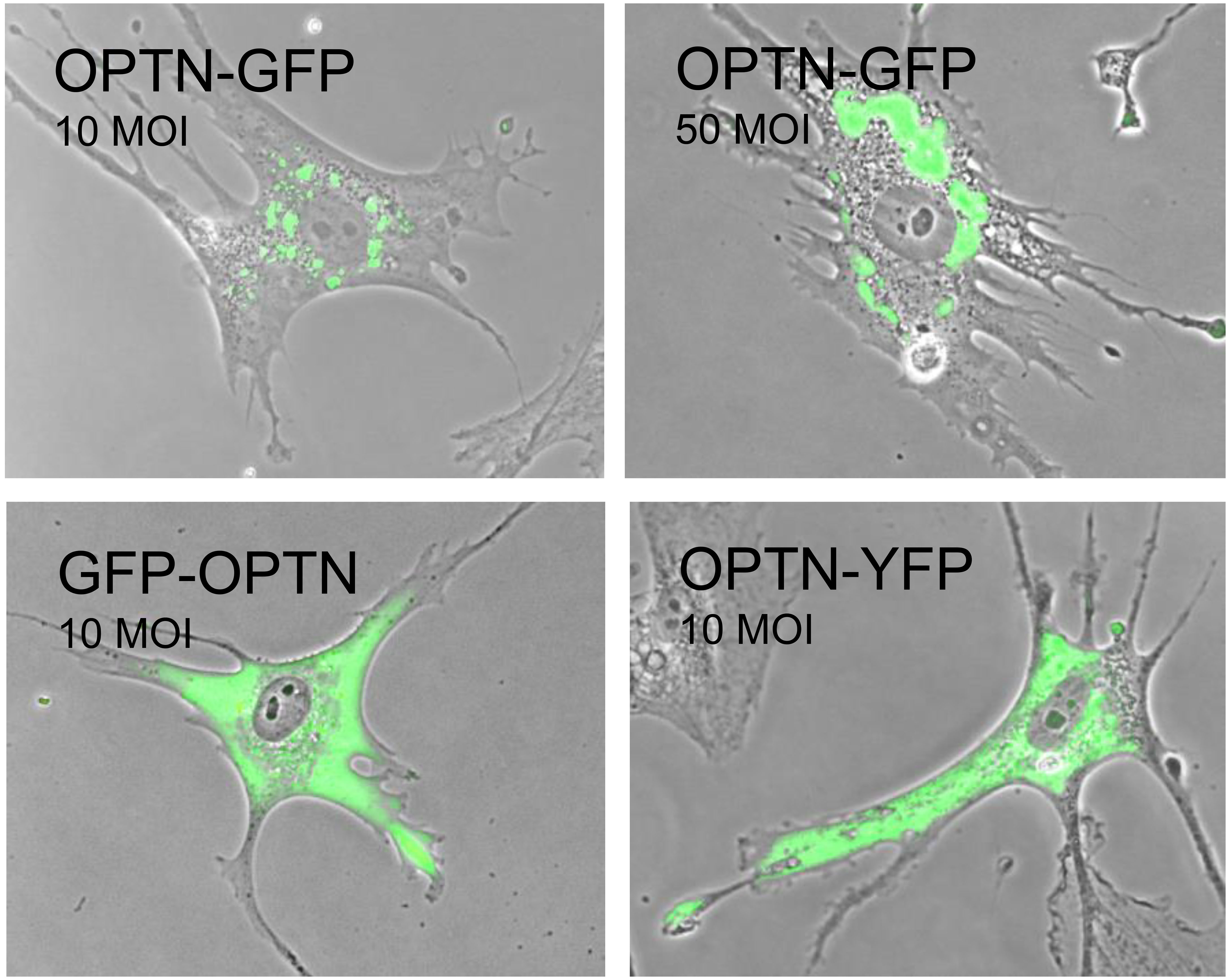
 Figure 4 of
Suh, Mol Vis 2024; 30:114-122.
Figure 4 of
Suh, Mol Vis 2024; 30:114-122.