Figure 1. En face view of the corneal endothelial transition zone (white arrow) between the corneal endothelium and trabecular meshwork
in different laboratory animals. Some cell clusters with two or three layers were identified; moreover, weaker Alizarin Red
staining and greater polymorphic cells were observed in rabbit (A), rat (B), and mouse (C) specimens. The scale bar indicates
100 μm.
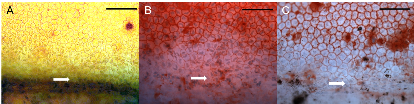
 Figure 1 of
Lee, Mol Vis 2024; 30:107-113.
Figure 1 of
Lee, Mol Vis 2024; 30:107-113.