Figure 12. SAL attenuated phosphorylation of NF-𝜅B p65 translocation to the nucleus by enhancing the PI3K/Akt/GSK-3β signaling pathway.
Immunofluorescence analysis of NF-𝜅B p65 translocation to the nucleus in rMC-1 cells. A: The nuclear translocation of NF-𝜅B p65 was determined by microscopy. B: Fifteen fields were imaged per well, and cells in which NF-𝜅B was located in the nucleus were counted. These data are expressed
as a ratio to the total number of cells in the field. The experiments were performed in triplicate, and the values are expressed
as the percentage of cells per field that localized NF-𝜅B to the nucleus. Values are presented as mean ± SD (n=3). Scale
bar=50 μm. **p<0.01, versus CON group. #p<0.05, versus HG group. △p<0.05, versus HG+SAL group.
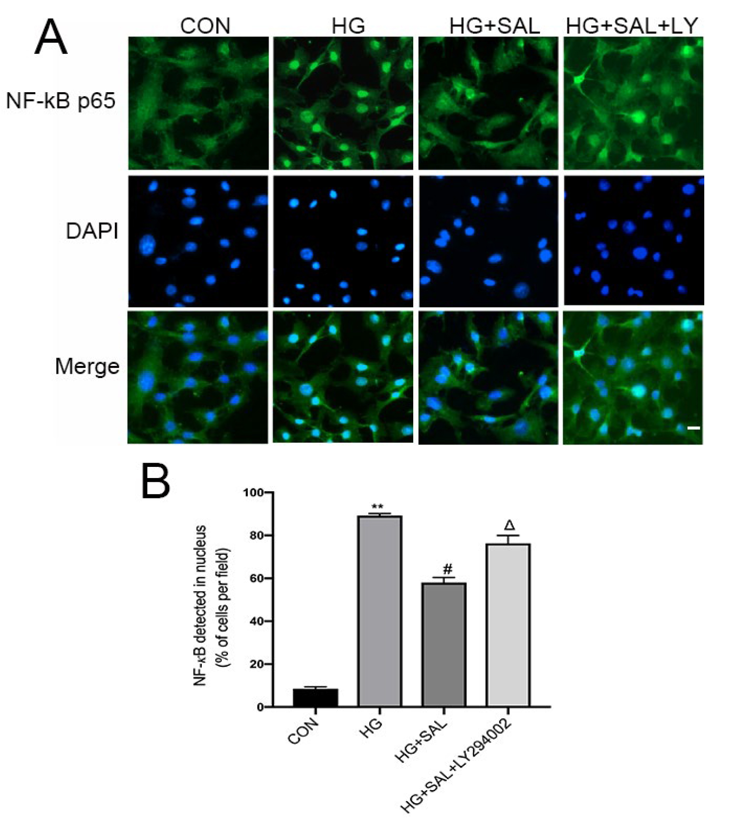
 Figure 12 of
Feng, Mol Vis 2024; 30:1-17.
Figure 12 of
Feng, Mol Vis 2024; 30:1-17.