Figure 1. SAL prevents ganglion cell loss due to STZ-induced injury. A: H&E staining showing that SAL protects ganglion cells in diabetic rats. The arrowheads represent retinal ganglion cells.
B: Quantitative analysis of the number of ganglion cells. SAL-treated retinas showed a significantly higher number of ganglion
cells than the DM group. (#p<0.05). Values are presented as mean ± SD (n=3). Scale bar=100 μm. ***p<0.001, versus CON group. #p<0.05, versus DM group.
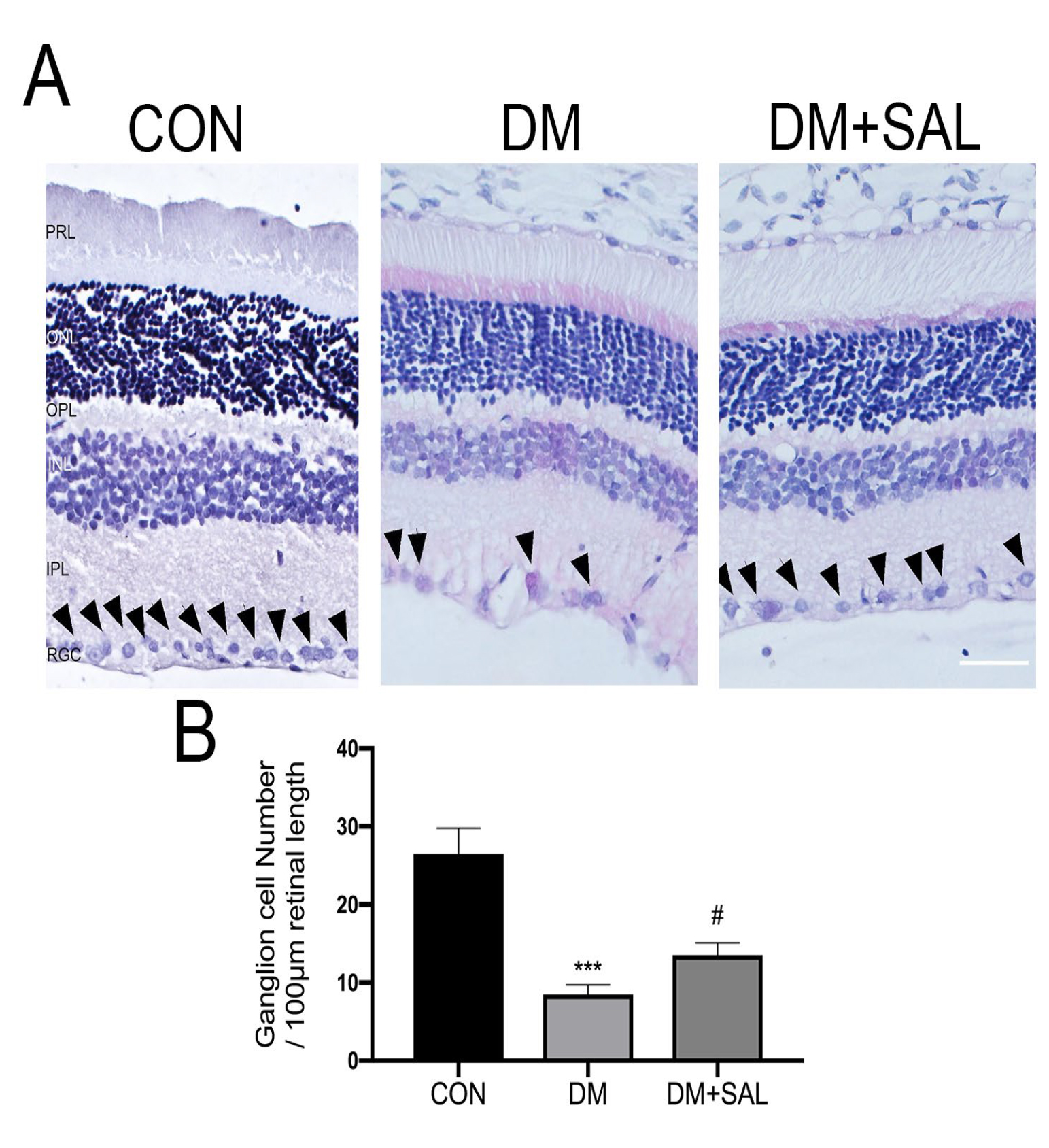
 Figure 1 of
Feng, Mol Vis 2024; 30:1-17.
Figure 1 of
Feng, Mol Vis 2024; 30:1-17.