![]() Figure 4 of
Segovia, Mol Vis 3:9, 1997.
Figure 4 of
Segovia, Mol Vis 3:9, 1997.
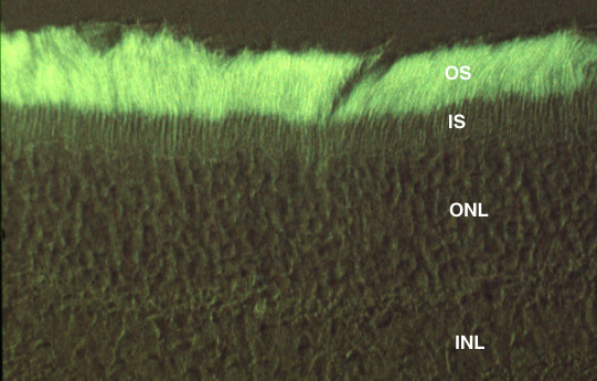
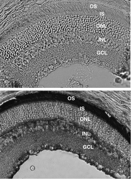
Figure 4. Localization of µ-crystallin in adult rat retina
(a) Immunohistochemistry using peroxidase staining. Above, rat retina with pre-immune serum. Below, similar section with µ-crystallin antiserum.
(b) Immunofluorescence using µ-crystallin antiserum. Retina layers are indicated; OS, photoreceptor (rod) outer segments; IS, photoreceptor inner segments; ONL, outer nuclear layer; INL, inner nuclear layer; GCL, ganglion cell layer.