![]() Figure 3 of
Segovia, Mol Vis 3:9, 1997.
Figure 3 of
Segovia, Mol Vis 3:9, 1997.
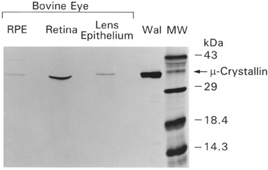
Figure 3. Detection of µ-crystallin in bovine eye tissues
Western blot of adult bovine eye tissues positive for µ-crystallin immunoreactivity; retinal pigment epithelium (RPE), retina and lens epithelium. Wal shows results for tammar wallaby (Macropus eugenii) lens protein as positive control. µ-Crystallin is detected at a size of 34 kDa. In this gel the separation of the major and minor µ-crystallin bands is closer than in Figure 1. Bovine tissues show only a single band. Other eye tissues, lens fibers, cornea, iris and sclera were negative (not shown).