![]() Figure 2 of
Shearer, Mol Vis 3:8, 1997.
Figure 2 of
Shearer, Mol Vis 3:8, 1997.
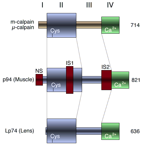
Figure 2. Domains of calpain isozymes
Schematic domain structures for m-calpain and µ-calpain compared to tissue-type calpains found in muscle (p94) and rat lens (Lp74). The Roman numerals designate domains I (autolytic), II (cysteine protease catalytic site), III (unknown function), and IV (calmodulin-like calcium binding regions) for the family of calpains. NS (novel sequence), IS1 (insert 1) and IS2 (insert 2) in red are sequences unique to p94 and deleted in Lp74. The ubiquitous m-calpain and µ-calpain also have domains I-IV, but are separate gene products showing 44-49% homology to p94 and Lp74. Numbers to the right are total amino acid residues in the primary sequences.