 Figure 6 of Dhawan et al
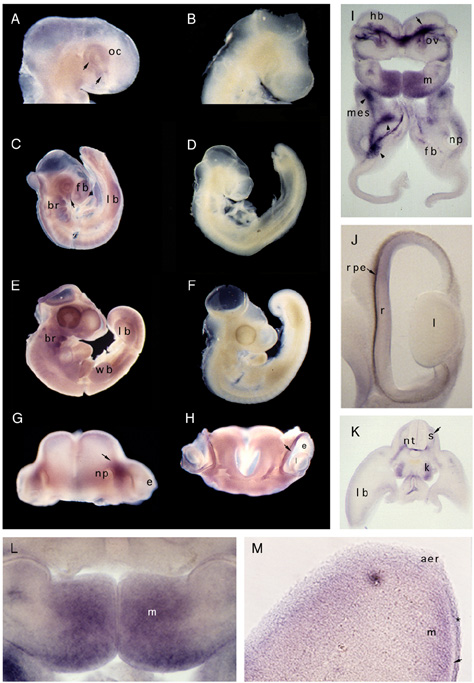
Figure 6. Whole mount in situ hybridization of chicken embryos, probed with digoxigenin labeled chdlx-3 riboprobes.
A, C, E, G, and H were probed with the anti-sense strand and B, D, and F were probed with the sense strand. The embryo in A shows dlx- transcripts in the optic cup (arrows) at early D3 (stage 16) of development. In C, at stage 18 of development, the chdlx-3 transcripts were present in the forebrain (fb), the optic cup (arrow), the branchial arches (br), and the leg bud (lb). At 3.5 days (stage 22, embryo in E), chdlx-3 transcripts were more abundant in the optic cup and the branchial arches. Staining is also seen in the limb buds (lb and wb). G is the anterior view of the frontal lobes and the nasal placode of a D3.5 embryo. Intense staining is seen in the mesenchyme in the region between (arrow) the eye (e) and nasal placode (np). Faint staining is also seen in the whole frontal lobe. H is the posterior view of the tissue in G, showing low levels of staining in the mesenchyme of the frontal lobes and more intense staining in the retina (arrow). I, J and K are 70 µm Vibratome sections of stage 22 embryo. I is through the floor of the hindbrain (hb), otic vesicle (ov), mandibular arch (m), the nasal placode (np), and the forebrain (fb). Intense staining is seen in the mesenchyme beneath the floor of the hindbrain (arrow), in the mandibular arch (m), and the cranial ganglia (arrow heads). J is a section through the right eye in H, showing staining in the retina. K is a transverse section through the trunk at the level of leg buds. Staining is seen in the migrating neural crest (arrow), in the mesonephros (k) and in the peripheral avascular zone of the mesoderm in the leg bud. L and M are higher magnification (20X) views of similar sections as in I and K. L shows that staining in the mandibular arch is restricted to the cells of the mesenchyme. In M localization of chdlx-3 transcripts in the dorsal ectoderm (*) and the peripheral avascular mesenchyme (m) of the limb is apparent. The unstained region under the dorsal ectoderm is clearly distinguishable (arrow). The apical ectodermal ridge (aer) was not stained appreciably.
Click on the figure below to bring up a much larger (374k) version.
Mol. Vis. 3:7, 1997 [http://www.emory.edu/molvis/v3/dhawan]
©1997 Molecular Vision
ISSN 1090-0535