![]() Figure 2 of Dhawan et al
Figure 2 of Dhawan et al
Figure 2. An agarose gel showing the PCR products generated from several ocular tissues.
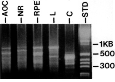
Tissues were isolated from stage 22 embryos, RNA was extracted, reverse transcribed and amplified with two sets of primers, as shown in Fig. 1. The pattern of bands obtained from each tissue was unique.
AOC, anterior optic cup; NR, presumptive neural retina; RPE, retinal pigment epithelium; L, lens; C, corneal epithelium; STD, molecular weight standards.