![]() Figure 5 of Kolb et al
Figure 5 of Kolb et al
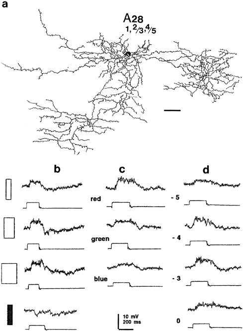
Figure 5:
Light reponses of A28 cell.
a) Camera lucida drawing of an intracellularly recorded and HRP labeled A28 reconstructed from the slice stain. Scale bar 50 um.
b) Light responses of the cell as revealed by intracellular recording and staining in the slice preparation. Responses to slits of various widths (from top to bottom: 0.025 mm; 0.45 mm and 0.66 mm slit width) and a black bar (0.15 mm width) surrounded by light. Log. rel. int. = -2.
c) Responses to 621 nm (red), 505 nm (green) and 404 nm (blue) monochromatic slits (0.45 mm width; 2.3 x 10⁸ Quanta/sec/cm2).
d) Responses to 621 nm slit stimuli (0.45 mm width) of increasing intensity. Log. relative intensity is indicated by numbers. Full intensity (Log. rel. int. = 0) was 2.3 x 10¹² Quanta/sec /cm2.