![]() Figure 4 of Kolb et al
Figure 4 of Kolb et al
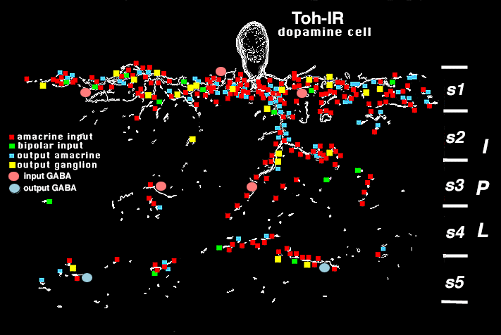
Figure 4:
Reconstruction of a Toh-IR cell body and main dendrites in S1 and passing to S2/3 and S4/5.
Other Toh-IR dendrites in the tissue were studied but did not necessarily get traced to the Toh-IR cell body. In the 15 um thick slab of tissue studied by EM, the Toh-IR cell and related processes received mostly amacrine synapses (red) in all strata of the IPL, and a few bipolar ribbon synapses (green) to the main tiers of dendrites in stratum 1, strata 2/3 and 4/5 borders. Toh-IR stained profiles were presynaptic to amacrine and ganglion cell processes (blue, yellow). Postsynaptic ganglion cell dendrites costratified in the three main dendritic tiers of the Toh-IR cell.