![]() Figure 3 of Kolb et al
Figure 3 of Kolb et al
Figure 3: EM of Toh-IR amacrine cells: Toh-IR profiles presynaptic to bipolar axons, ganglion cell
dendrites and amacrine cell profiles
(Click on any figure to see an expanded version. Caution: Expanded figures are 100K+)
| a) Toh-IR processes are presynaptic to other Toh-IR processes (white arrow) and to a ganglion cell (GC) dendrite (black/white arrow) in S1. The GC dendrite is also postsynaptic to an unstained amacrine cell (A). A neighboring unstained amacrine cell (A) is presynaptic to another Toh-IR process. | 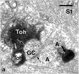
|
| b) A vesicle-filled Toh-IR process is presynaptic (black/white arrow) to an unstained amacrine cell (A) in S1 of the IPL. | 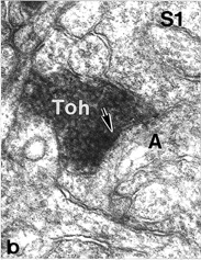
|
| c) A large Toh-IR process makes a synapse upon another smaller Toh-IR profile in S1. | 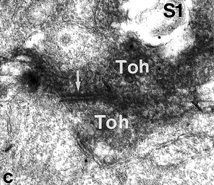
|
| d) A Toh-IR profile on the S4/5 border synapses upon a colloidal gold-containing GABA-IR (Ga) amacrine profile. Scale bar 0.5 um. | 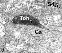
|