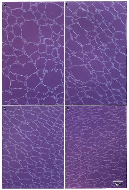
A. Embryonic Nucleus. The primary fiber cells in
humans are quite similar to bovine primary fibers in both size and
arrangement. Location = center of the equatorial plane.
B-F. Fetal
nucleus. In the fetal nucleus (B-E), considerable reduction in cross-
sectional size is evident as regions further from the lens center are
examined. The rounded profiles (B-C) gradually become flattened and
irregular (D-F), but do not have the hexagonal shape seen in bovine lenses.
Short rows of cells (C) become organized into radial cell columns (E),
however, these are more difficult to discern in the human than in the
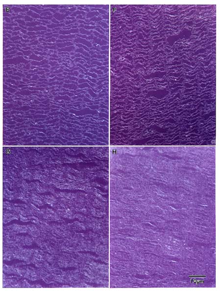
bovine. The fibers continue to flatten (F) and the profiles take on an
undulating appearance which is also seen in panels G and H.
B. Location =
0.6 mm from the lens center.
C. Location = 1 mm from the lens center.
D.
Location = 1.3 mm from the lens center.
E. Location = 1.5 mm from the
lens center.
F. Location = 2 mm from the lens center.
G. Juvenile
Nucleus. Two distinct cell sizes are characteristic of the juvenile nuclear
region, where larger cells are interspersed among the highly flattened cell
profiles. Location = 2.4 mm from the lens center.
H. Adult Nucleus. Cells
of the adult nuclear region are highly compressed, making their profiles
difficult to distinguish. Location = 3.5 mm from the lens center.