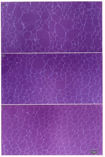
A. Embryonic nucleus. The largest cells are found in the
embryonic nucleus, where the disordered packing and large range in cross-
sectional area is apparent. Location = center of the equatorial plane.
B-D.
Fetal Nucleus. Panels B-D display how the cell size, shape and
arrangement gradually changes from disordered, rounded profiles which
vary greatly in size (B), to flattened hexagonal profiles of more uniform
size (D). Short irregular rows are discernable in C, which become easily
recognizable radial cell columns in D.
B. Location = 0.6 mm from lens
center.
C. Location = 1 mm from the lens center.
D. Location = 2 mm from
the lens center.
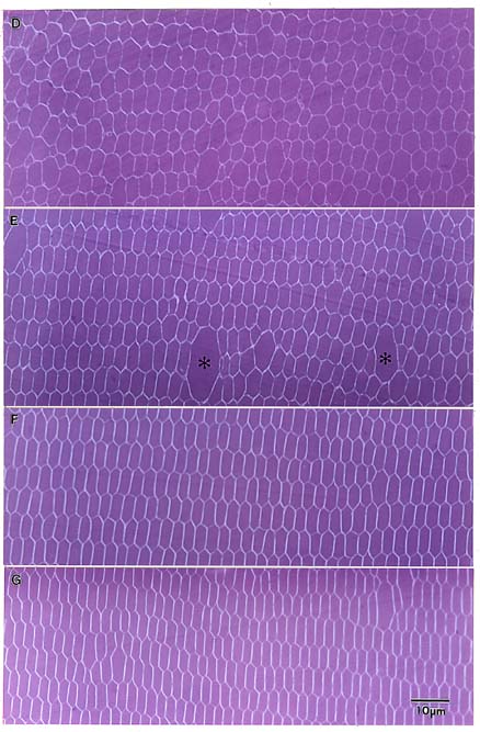
E. Juvenile Nucleus. The juvenile nuclear region is
characterized by occasional large profiles interspersed among relatively
uniform fiber cells arranged in regular radial cell columns. Location = 3
mm from the lens center.
F-G. Adult Nucleus. Fiber cells of the adult
nucleus (F-G) are highly flattened hexagons in shape and display very
regular packing. Cell profiles in figure G (located 5 mm from the lens
center) appear more flattened than those in figure F (located 4 mm from
the lens center.