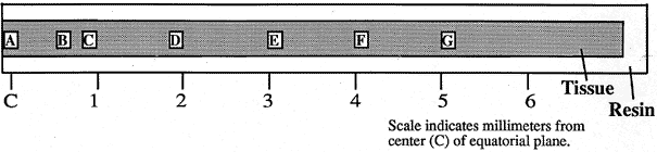
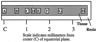
A. Vibratome sectioning of lenses. Lenses were mounted and sectioned parallel to the optic axis. A = anterior pole , P = posterior pole. Vibratome sections at or near the optic axis contained all the developmental regions of the lens: c = cortex, a = adult nucleus, j = juvenile nucleus, f = fetal nucleus, e = embryonic nucleus. Vibratome sections were bisected along the equatorial plane and thick sections (0.5”m to 1.0”m) were cut along 1/2 of the equatorial axis, stained with toluidine blue, and mounted on glass slides for light microscopic examination.