![]() Figure 4 of
Ban, Mol Vis 3:18, 1997.
Figure 4 of
Ban, Mol Vis 3:18, 1997.
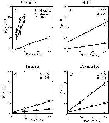
Figure 4. Flux of tracers across RPE monolayers.
The flux of HRP, mannitol and inulin across the RPE monolayer was linear for 90 min. (A) Each solute diffused rapidly across control filters that lacked cells. The rate of diffusion decreased with increased size of the tracer. A lag was noted for the diffusion of HRP that was minimized by preincubating the filters in HRP, as described in Methods (B,C,D). Three to four cultures were established from E10 embryos, and maintained in SF2 (open symbols) or stimulated with E14 retinal conditioned medium (closed symbols). After 9 days in culture, the flux of HRP (B), [3H]inulin (C) and [3H]mannitol (D) was measured in the apical to basal direction. Standard error bars were sometimes smaller than the symbol, and linear regression lines are included in panels B, C and D. RPE monolayers retarded the diffusion into the basal chamber. Cultures stimulated with E14 retinal conditioned medium had lower permeability. Similar results were obtained with E7 and E14 cultures and are summarized in Figure 5.