![]() Figure 9 of
Lin, Mol Vis 3:17, 1997.
Figure 9 of
Lin, Mol Vis 3:17, 1997.
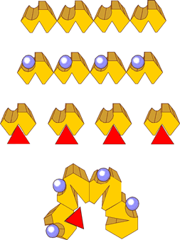
Figure 9. The positions of binding sites in IRBP.
This figure represents a model for the distribution of retinol (depicted as red triangles) and 16-AP (blue spheres) binding sites in IRBP (shown in yellow). We propose that there are four binding sites for retinol, with one in Domain B of each repeat. The top line depicts IRBP without any ligands bound. The next line down depicts IRBP with 16-AP bound. The binding of 16-AP to one site does not affect the binding properties of other 16-AP binding sites. The third line depicts individual repeats with retinol bound, showing that each repeat can bind one retinol molecule. The bottom line depicts IRBP with one retinol bound to one repeat, we speculate that a steric change might prevent other retinol ligands from binding to IRBP. The bent form may possess a lower affinity for additional retinoids, while the individual repeats may possess higher affinities because they are not bent. Alternatively, the structural change might affect the fluorescence enhancement of the additional sites in bent wild type protein. The pictured bent form of IRBP is based on the structural transition identified by Adler et al. (46) that accompanies retinol binding in bovine IRBP. With retinol bound or not, the 16-AP binding properties may remain unaffected. Further experiments with variant proteins should to resolve fundamental questions of how the Visual Cycle works.