![]() Figure 8 of
Lin, Mol Vis 3:17, 1997.
Figure 8 of
Lin, Mol Vis 3:17, 1997.
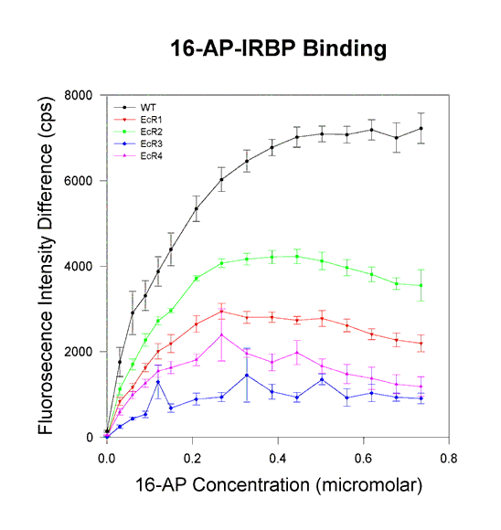
Figure 8. Ligand binding assays: 16-AP binding to altered IRBP proteins.
One micromolar protein was used in each sample. The concentrations of the ligand are shown on the abscissa, and the fluorescence difference between the sample (protein plus 16-AP) and the blank (16-AP only) in photons counted per second (cps) is shown on the ordinate. The raw data from 16-AP titrations of the WT insect cell-derived protein and from E. coli produced individual repeats proteins are shown. The results from several independent assays are averaged. The error bars indicate the standard errors of the mean. WT protein (in black) shows the highest binding followed by EcR2 in green, EcR1 in red, EcR3 in blue, and EcR4 in violet.