![]() Figure 1 of
Lin, Mol Vis 3:17, 1997.
Figure 1 of
Lin, Mol Vis 3:17, 1997.
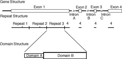
Figure 1. IRBP gene structure.
The relationship among IRBP gene structure, repeats in the protein, and domains within the repeats (22, 66). The line drawings are roughly to scale. The top line represents the IRBP gene exons and introns illustrating the very long first exon and the normal sizes of the remaining exons. The middle line drawing shows the four protein repeats. The bottom line shows the two putative domains within each of the repeat units. Two complex carbohydrate attachment sites are found in human IRBP. Their locations are marked by the asterisks. Based on homology with Tsp, in which active site residues have been identified, we suggest that a hydrophobic retinol binding site resides in Domain B. By analogy to other proteins in the family, Domain A may represent a regulatory domain, analogous to PDZ or PTB domains (67). Each repeat shows evidence of possessing an all-trans-retinol and 16-AP binding site in the human IRBP protein, as detailed in this report.