![]() Figure 5 of
Ostrer, Mol Vis 3:16, 1997.
Figure 5 of
Ostrer, Mol Vis 3:16, 1997.
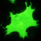
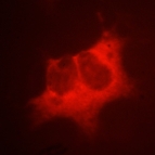
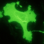
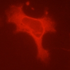
Figure 5. Immunofluorescence analysis of 293-EBNA cells expressing Grn and P307L
Cells were incubated with either the 1D4 mAb and a secondary fluoresceinated goat anti-mouse Ab (opsin) or with RIC6 and RIIL3 polyclonal antibodies and a secondary Texas red-conjugated goat anti-rabbit Ab (ER). No differences were observed in the immunofluorescence patterns of the wild type and mutant opsins, indicating that the mutant opsin is transported normally to the cell membrane.
 |  | |
 |  |