![]() Figure 4 of
Ostrer, Mol Vis 3:16, 1997.
Figure 4 of
Ostrer, Mol Vis 3:16, 1997.
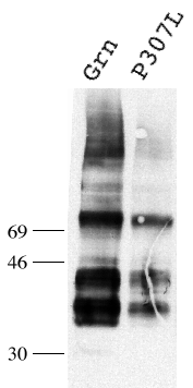
Figure 4. Western blot analysis of wild type and mutant green opsins
Polypeptides from lysates of equal numbers of cells were separated by 12% SDS-PAGE, transferred to nitrocellulose, and probed with 1D4 mAb. Immunoreactive bands were visualized using chemiluminescence. The band just below the 46 kd molecular weight marker represents the fully glycosylated form. All bands above 46 kd represent multimeric forms of the protein. No differences were observed in the glycosylation patterns of the wild-type and mutant opsins.