![]() Figure 1 of
Ostrer, Mol Vis 3:16, 1997.
Figure 1 of
Ostrer, Mol Vis 3:16, 1997.
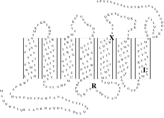
Figure 1. A secondary structure model of green cone opsin
The location of the P307L mutation is highlighted. The locations of other mutations found in blue cone monochromats are also shown. The amino acid sequence of the 1D4 epitope is shown in bold. "R" indicates the substitution of arginine for cysteine at position 203. "X" indicates the substitution of a premature terminator for arginine at position 247.