![]() Figure 11 of
Boatright, Mol Vis 3:15, 1997.
Figure 11 of
Boatright, Mol Vis 3:15, 1997.
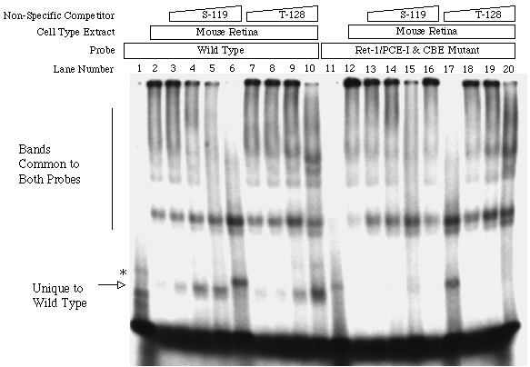
Figure 11. EMSA of wild-type and mutant activator sequence probing against extracts from whole mouse retina.
This experiment was conducted identically to the previous experiment (Figure 10), except extracts probed were from mouse retina tissue rather than chick retinal cell cultures; results are similar. Both probes (the -70 to -45 sequence of the murine IRBP promoter, with or without base transversions in Ret-1/PCE-I and the CRX-binding element [CBE]) bind many complexes with similar migration patterns. The wild type probe binds a complex (arrow) that appears just below the artifactual band (asterisk) seen in lanes with probe alone (lanes 1 and 11) and in lanes where protein binding is precluded by high concentrations of dispersant.