![]() Figure 4 of
Evans, Mol Vis 3:11, 1997.
Figure 4 of
Evans, Mol Vis 3:11, 1997.
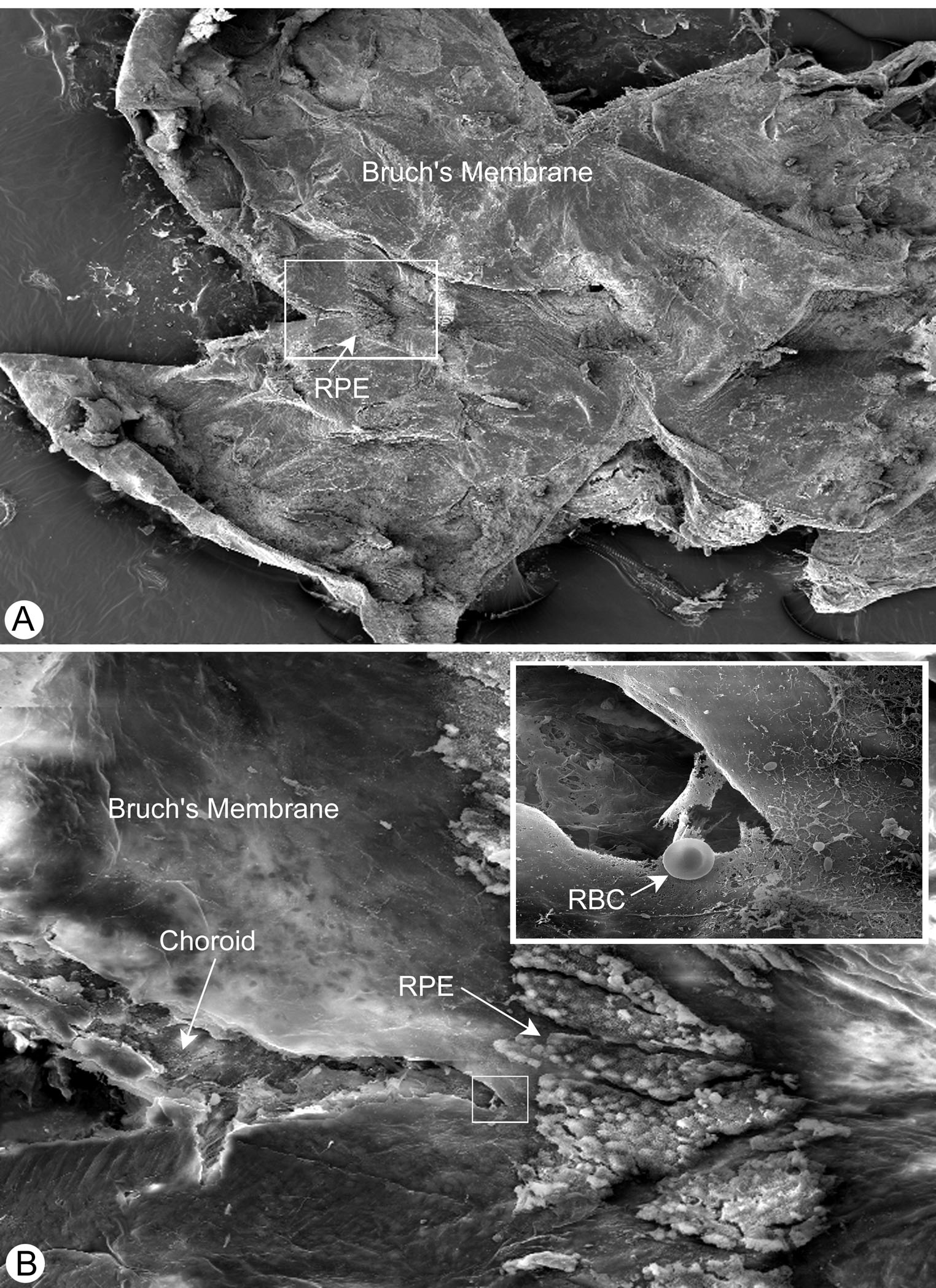
Figure 4. Scanning electron photomicrograph of a vitiligo mouse eye cup demonstrating that samples obtained for the esterification assays were enriched for RPE.
(A) A 9 week old vitiligo mouse eye cup seen at 28X magnification, showing a few patches of remaining RPE cells and a largely intact Bruch's membrane after gentle brushing with a small artists brush.
(B) An enlargement to 225X magnification of the boxed area in (A) reveals a patch of RPE cells and an intact Bruch's membrane. A small portion of choroid is revealed following clipping with dissecting scissors when the eye cup is laid flat during brushing. The inset, at 3628X magnification reveals minimal disruption to Bruch's membrane and the underlying choroid.