Figure 7. Rod outer segment (OS) phagocytosis 1 day after stencil removal. OS (bright green) were seen in all samples, but fewer appeared
near the edge where junctional proteins were affected. Cells grown without a stencil that did not experience detachment were
used as controls. Scale bars: 50 µm.
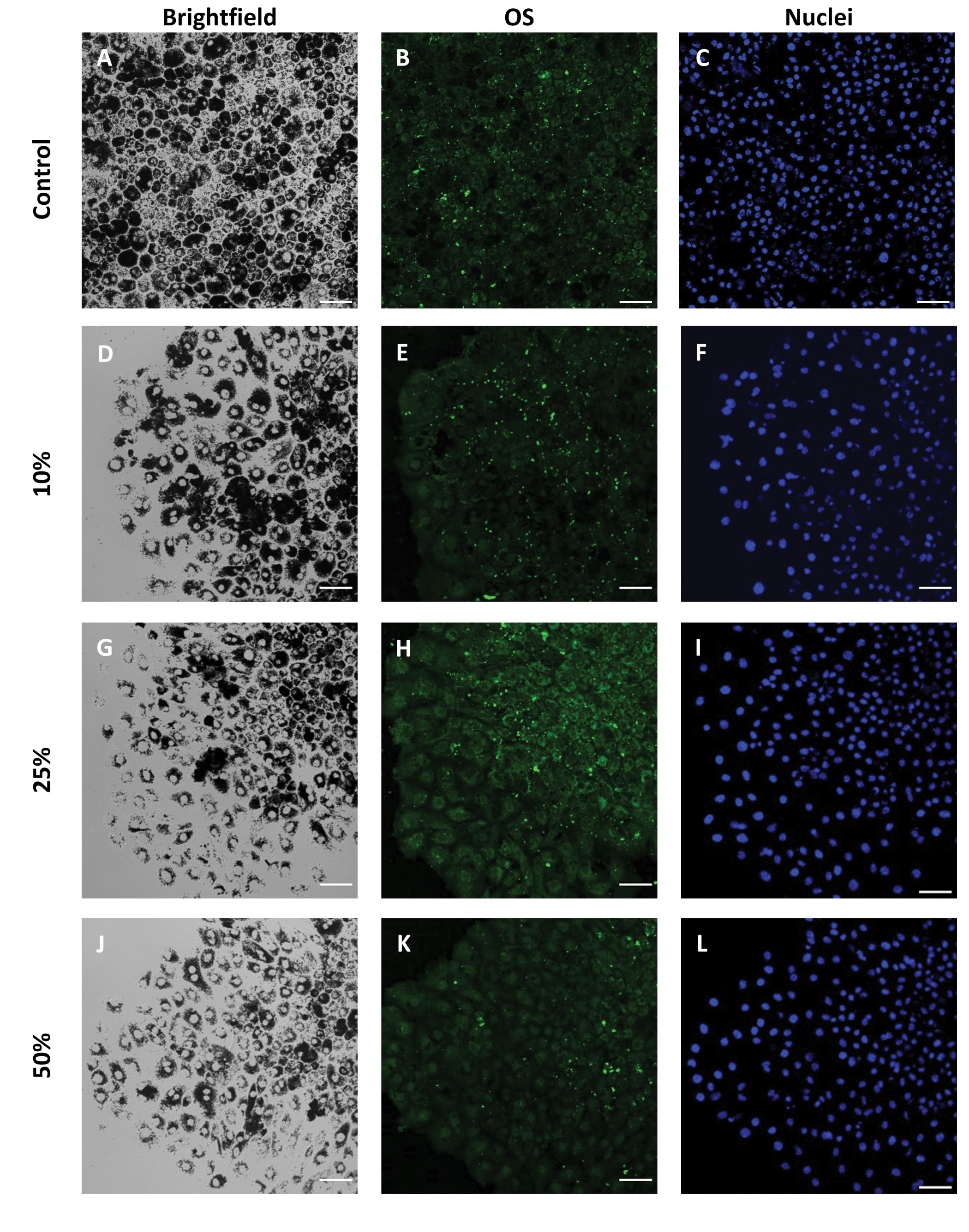
 Figure 7 of
Paterson, Mol Vis 2023; 29:87-101.
Figure 7 of
Paterson, Mol Vis 2023; 29:87-101.