Figure 8. Comparison of rabbit corneas at different time points after Photorefractive Keratectomy surgeries. A, B: Triplex IHC for vimentin, SMA, and nidogen-1 in corneas at different time points after PRK surgeries. Triplex IHC for mesenchymal
marker vimentin (green), myofibroblast marker alpha-SMA (red), and epithelial BM marker nidogen-1 (magenta) was observed in
rabbit corneas at different time points after −3D PRK. At 1 and 2 weeks after PRK, there was a patchy expression of nidogen-1
with irregular cellular arrangement of epithelial cells. There was evidence for few vimentin-positive fibroblasts at both
of these time points, and vimentin-positive fibrocytes could also be present in the anterior stroma. At 4 weeks after PRK,
there was an increase in the number of vimentin-positive cells in the anterior stroma. In parallel, there was an increase
in the percentage of epithelial BM regenerated to around 80–90%. This suggests a strong association of fibroblasts with epithelial
BM regeneration at 4 weeks after PRK injury. At 6 weeks after PRK, when the percentage of epithelial BM regeneration was further
increased, the corneal fibroblast numbers had decreased. At 8 weeks after PRK, most vimentin-positive cells disappeared from
the anterior stroma, when the epithelial BM was fully regenerated. No alpha-SMA-positive cells were observed at any of the
time points tested, indicating there was no fibrosis in these corneas. Blue is DAPI staining of the nuclei. Mag.: 200×.
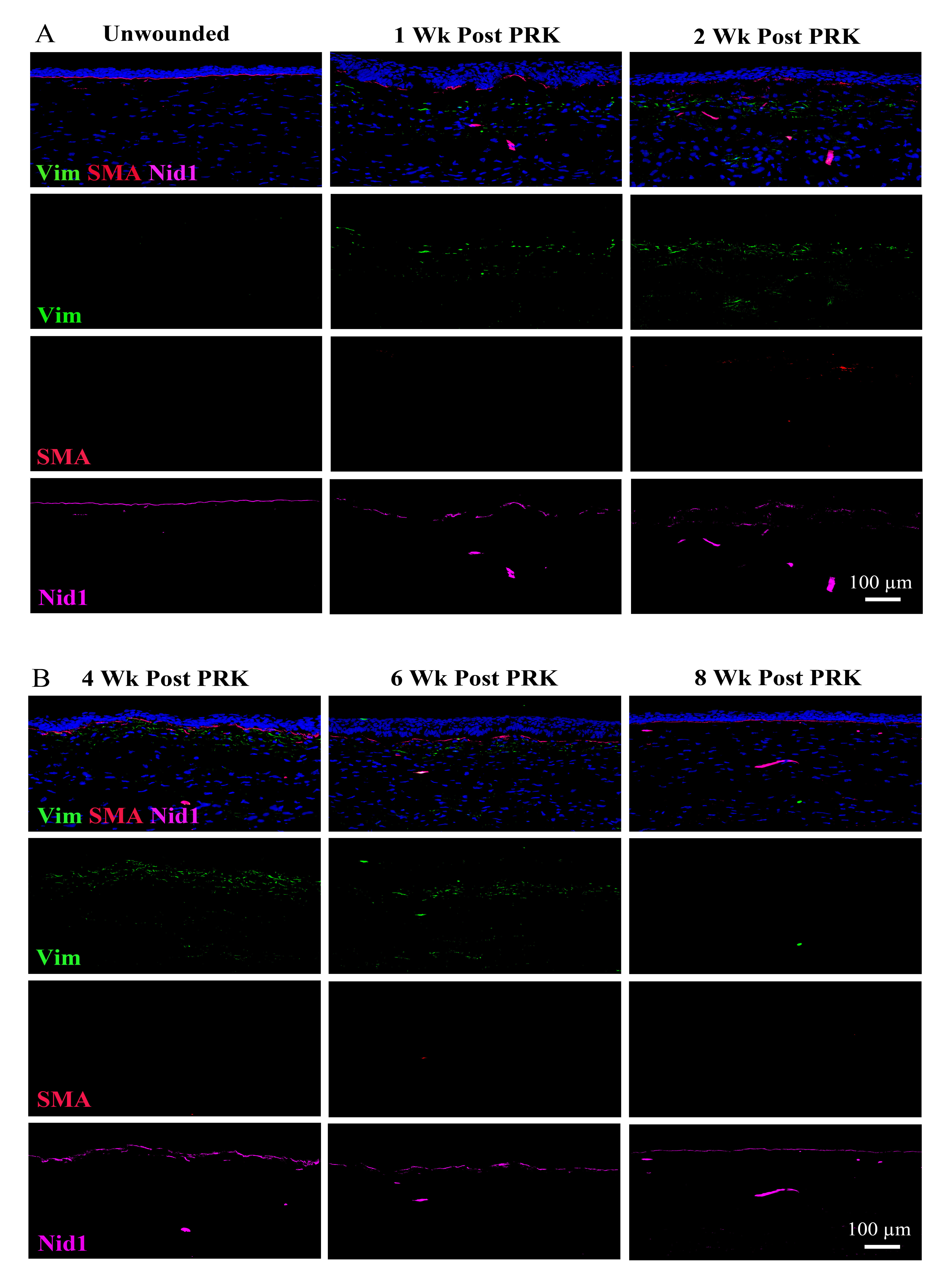
 Figure 8 of
Shiju, Mol Vis 2023; 29:68-86.
Figure 8 of
Shiju, Mol Vis 2023; 29:68-86.