Figure 6. IHC for laminin alpha-5, perlecan, and nidogen-1 in CMyo + Epi organotypic cultures. Triplex IHC did not detect epithelial
BM posterior to the epithelium in this example culture or any other corneal myofibroblast, bone marrow-derived myofibroblast,
or mixed corneal-bone marrow myofibroblast organotypic culture with corneal epithelium. Also, no stratified epithelium was
detected in these cultures. This indicates that the myofibroblasts derived from both cornea and bone marrow—either alone as
CMyo (100%)/BMyo (100%) or combined (CMyo 50% + BMyo 50%)—when incubated with corneal epithelial cells could not assemble
BM. Rather, spheroid-like epithelial masses were present in many cultures. Myofibroblasts were observed throughout the matrix.
Blue is DAPI staining of the nuclei. Mag.: 200×.
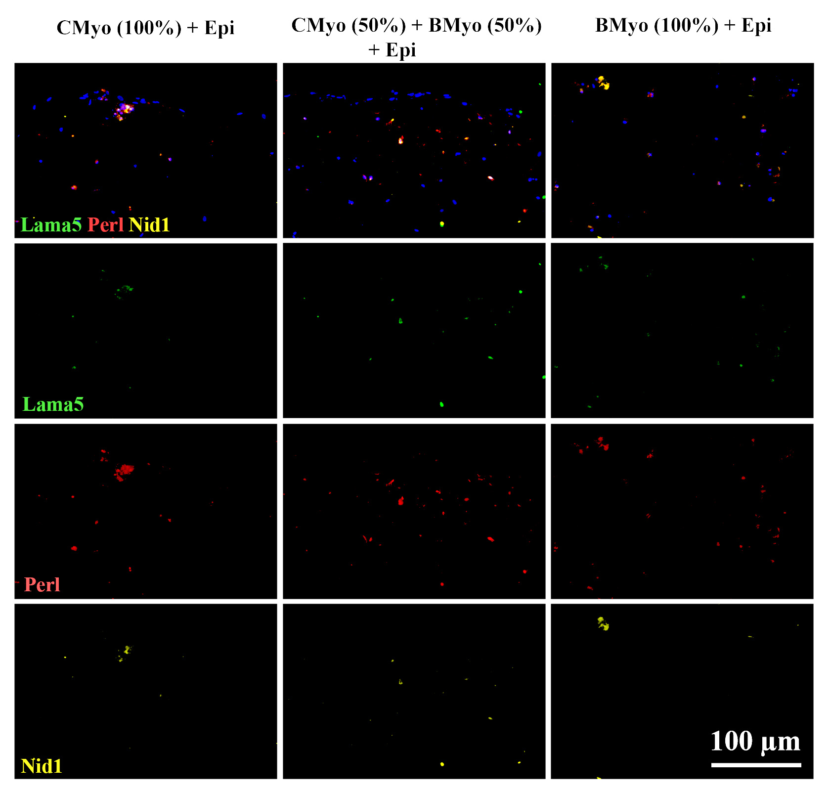
 Figure 6 of
Shiju, Mol Vis 2023; 29:68-86.
Figure 6 of
Shiju, Mol Vis 2023; 29:68-86.