Figure 4. Comparison of laminin beta 3 and collagen IV expression in different 3D cultures. A: Duplex IHC for laminin beta-3 and collagen type IV in corneal organotypic cultures. The CFib + Epi cultures showed high
expression of laminin beta-3 (green) in basal and more superficial epithelial layers. Collagen type IV (red) at the interface
of the epithelial cells and fibroblasts indicates a normally regenerated epithelial BM. However, CMyo + Epi cells in organotypic
cultures showed no evidence for epithelial BM generation, although myofibroblasts were found to produce collagen type IV.
In Epi only controls, laminin beta-3 is expressed throughout the epithelium, with low expression of collagen type IV. Blue
is DAPI staining of cell nuclei. Mag.: 200×. B: Graph showing percentage formation of laminin beta-3 and collagen IV compared between the groups of 3D cultures. Error bars
represent mean ± SEM. One-way ANOVA was used to compare between the groups, and p < 0.05 was considered statistically significantly
different.
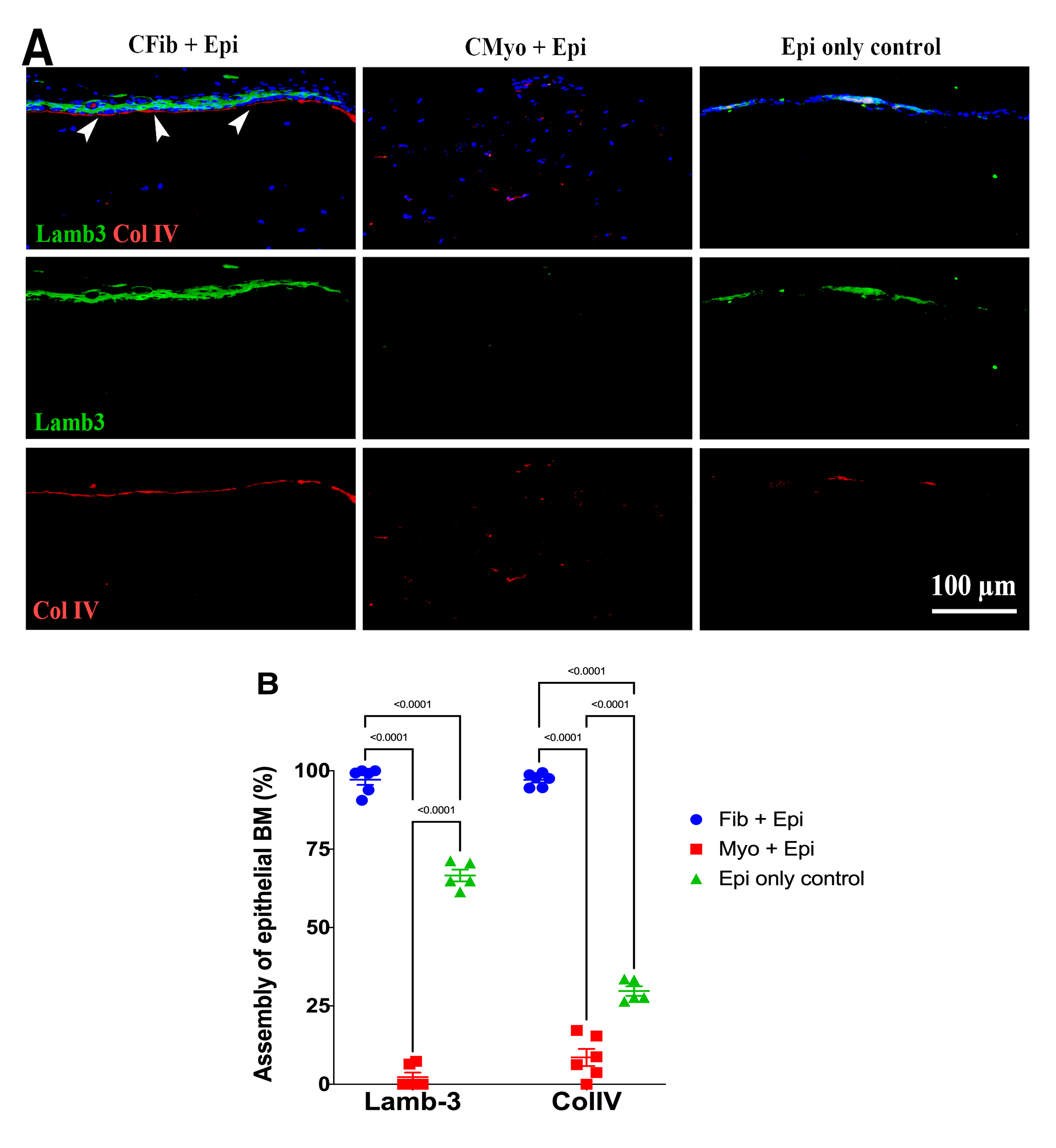
 Figure 4 of
Shiju, Mol Vis 2023; 29:68-86.
Figure 4 of
Shiju, Mol Vis 2023; 29:68-86.