Figure 3. Comparison of laminin alpha 5, perlecan and nidogen-1 expression in different 3D cultures. A: Triplex immunohistochemistry (IHC) for laminin alpha-5, perlecan, and nidogen-1 in 3D corneal organotypic cultures. The
corneal fibroblast and epithelial (CFib + Epi) organotypic cultures showed expression of laminin alpha-5 (green), perlecan
(red), and nidogen-1 (yellow) at the interface between the epithelial cells and the fibroblasts, suggesting a well-assembled
epithelial BM. In corneal myofibroblast and epithelium (CMyo + Epi) cultures, there was a high expression of perlecan, a lower
expression of laminin alpha-5, and no expression of nidogen-1 detected. It is important to note that the expression was observed
only in the stroma, and the overlying epithelial BM was not assembled when myofibroblasts were co-cultured with epithelial
cells. When the epithelial cells were cultured alone above the collagen matrix (Epi only control), laminin alpha-5 was predominantly
expressed throughout the epithelium, perlecan expression was low, and no expression of nidogen-1 was detected. Blue is DAPI
staining of cell nuclei. Mag.: 200×. B: A graph showing the percentage assembly of individual epithelial BM components, comparing between the groups of 3D cultures.
Error bars represent mean ± SEM. Each BM component was compared between the groups using one-way ANOVA, and p < 0.05 was considered
statistically significantly different.
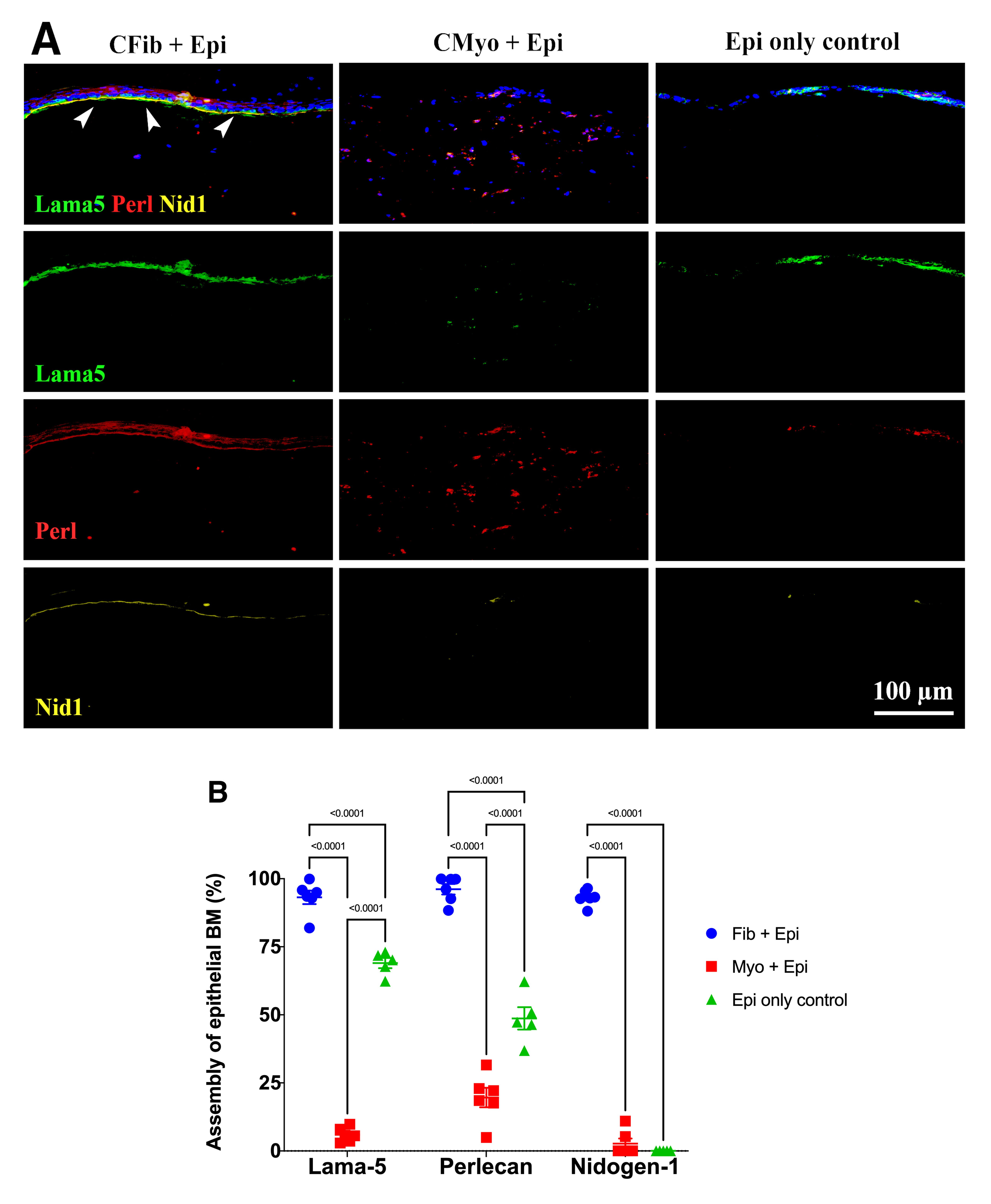
 Figure 3 of
Shiju, Mol Vis 2023; 29:68-86.
Figure 3 of
Shiju, Mol Vis 2023; 29:68-86.