Figure 1. Confirmation of cell morphology using light microscopy and immunocytochemistry A: Phase-contrast microscopy of rabbit corneal fibroblasts, myofibroblasts, and epithelial cells. Mag.:100×. B: Immunocytochemistry (ICC) for myofibroblast markers in cornea- and bone marrow-derived myofibroblasts. Myofibroblasts cultured
in transforming growth factor (TGF) beta-1 for 14 days had ICC for vimentin, alpha-smooth muscle actin (SMA), desmin, and
vinculin. Both cornea- and bone marrow-derived cells expressed vimentin, alpha-SMA, desmin, and vinculin, indicating they
were vimentin-alpha smooth muscle actin-desmin (VAD)-positive myofibroblasts (Chaurasia et al., 2009). ICC with the corresponding
isotypic control antibodies was also performed for each marker. Blue is 4′,6-diamidino-2-phenylindole (DAPI) staining of the
nuclei. Mag.: 200×.
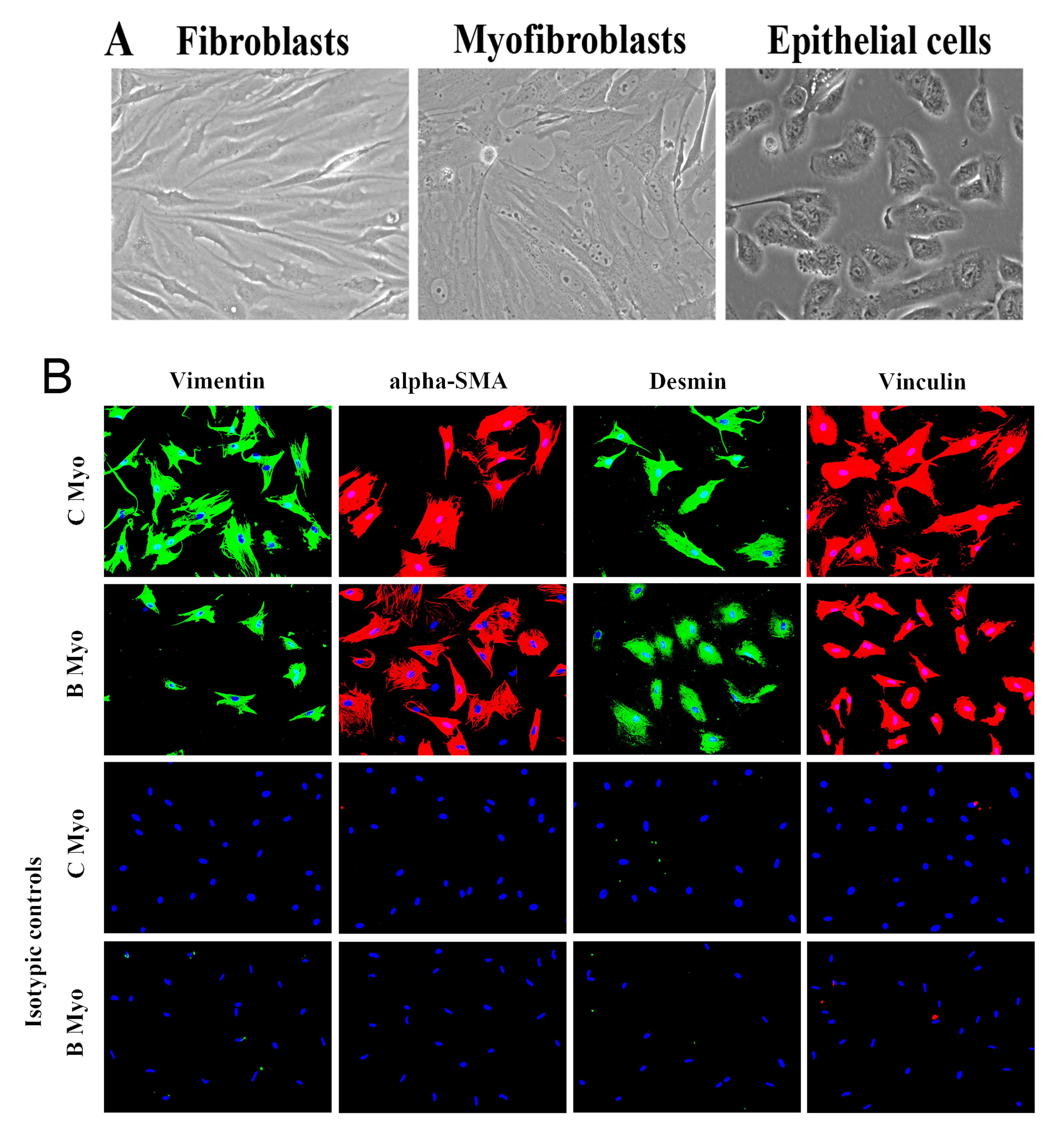
 Figure 1 of
Shiju, Mol Vis 2023; 29:68-86.
Figure 1 of
Shiju, Mol Vis 2023; 29:68-86.