Figure 6. Descemet’s membrane (DM) regeneration at different time points in rabbit corneas. A: In the normal cornea, DM was thick and
even (scale bar: 2μm, magnification: ×10,000). B: At 2 months after injury, a thin DM first appeared in the posterior stroma
near the nascent endothelium (scale bar: 500 nm, magnification: ×20,000). C: At 3 months after injury, thicker but uneven
DM appeared in the posterior stroma adjacent to the endothelium. (en) represents the endothelial cell. (D) indicates Descemet’s
membrane.
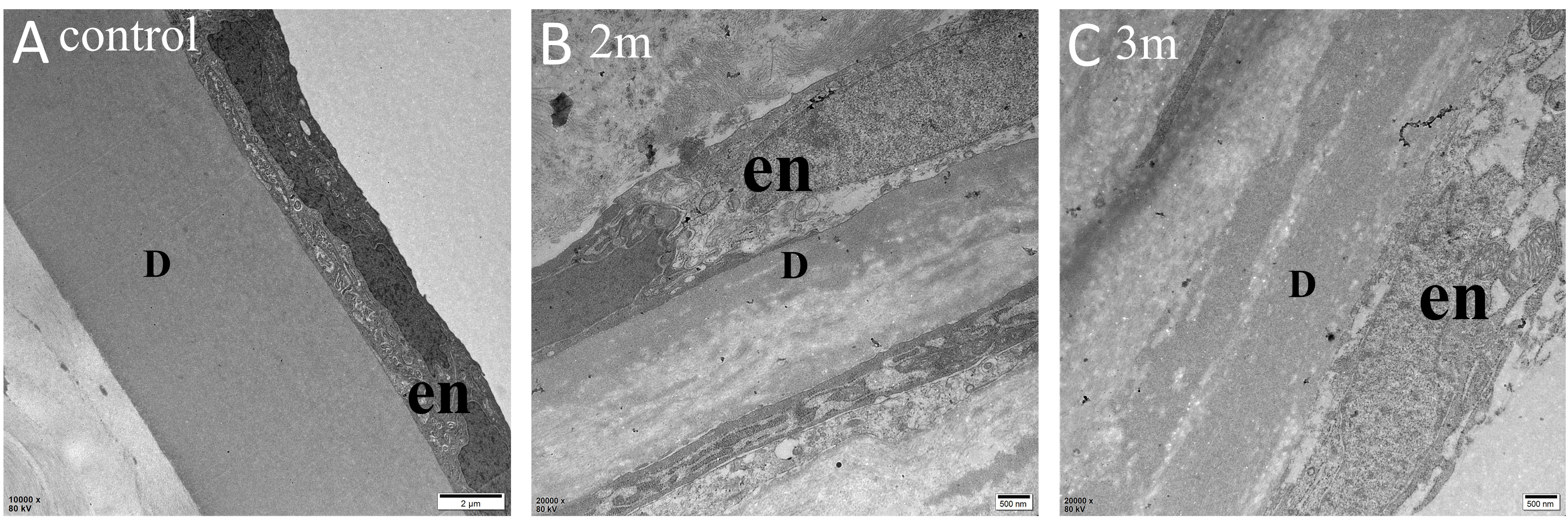
 Figure 6 of
Meng, Mol Vis 2023; 29:58-67.
Figure 6 of
Meng, Mol Vis 2023; 29:58-67.