Figure 5. Epithelial basement membrane (EBM) regeneration at different time points in rabbit corneas (scale bar: 500 nm, magnification:
×30,000). A: Unwounded (control) corneas had normal EBM ultrastructure with lamina lucida and lamina densa. B–D: After perforating
injury, all corneas at 3 days to 2 weeks showed no evidence of regeneration of lamina lucida and lamina densa. E: Three weeks
after injury, a nascent defective EBM was first observed, and the lamina lucida and lamina densa were irregular and discontinuous.
F: One month after injury, the EBM ultrastructure was somewhat restored, but its ultrastructural morphology was still uncontinuous.
G: Two months after injury, the EBM lamina lucida and lamina densa showed more continuity, and there were a few discontinuous
EBM ultrastructures. H: Three months after injury, continuous and regular lamina lucida and lamina densa were noted, and EBM
was nearly fully regenerated. Arrows indicate the lamina densa and lamina lucida of the epithelial basement membrane. A represents the control cornea. B–H represent injured corneas. (E) is the epithelium. (S) is the stroma.
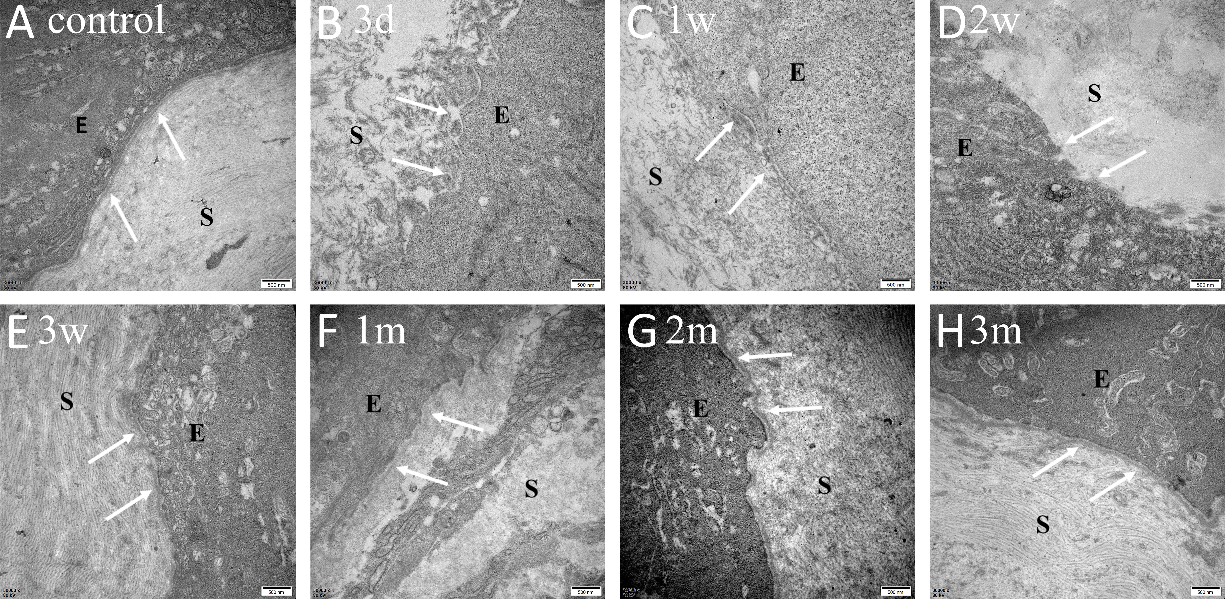
 Figure 5 of
Meng, Mol Vis 2023; 29:58-67.
Figure 5 of
Meng, Mol Vis 2023; 29:58-67.